9VGW | pdb_00009vgw

Crystal structure of human PCNA in complex with REV1 PIP-box


Experimental Data Snapshot

  • Method: X-RAY DIFFRACTION
  • Resolution: 2.70 Å
  • R-Value Free: 
    0.261 (Depositor), 0.260 (DCC) 
  • R-Value Work: 
    0.214 (Depositor), 0.214 (DCC) 
  • R-Value Observed: 
    0.216 (Depositor) 

Starting Model: experimental
View more details

wwPDB Validation 3D Report Full Report

Validation slider image for 9VGW

This is version 1.1 of the entry. See complete history

Literature

Identification of a PCNA-binding motif in human translesion DNA polymerase REV1 and structural basis of its interaction with PCNA.

Hishiki, A.Hoshino, N.Okawara, K.Fuchigami, S.Hara, K.Hashimoto, H.

(2025) J Biochem 178: 315-324

  • DOI: https://doi.org/10.1093/jb/mvaf054
  • Primary Citation Related Structures: 
    9VGW

  • PubMed Abstract: 

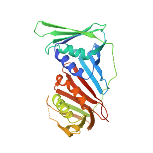
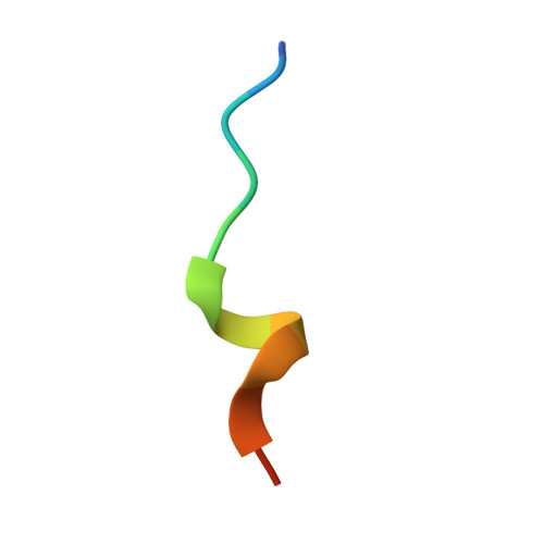
    REV1 is a eukaryotic error-prone DNA polymerase belonging to the Y-family, with a central role in translesion DNA synthesis (TLS) to continue DNA replication even in the presence of DNA damage in the template strand. TLS is stimulated by mono-ubiquitination of proliferating cell nuclear antigen (PCNA), a toroidal-shaped protein functioning as a scaffold for DNA polymerases and repair enzymes. Mammals possess four types of Y-family DNA polymerases: Pol η, Pol κ, Pol ι, and REV1. Among those, Pol η, Pol κ, and Pol ι interact with PCNA through PCNA-binding motifs, low-affinity variants of PCNA-interacting protein box (PIP-box). To date, several studies have reported that REV1 interacts with PCNA, but identified PCNA-binding regions are inconsistent; therefore, a structural basis for interaction between REV1 and PCNA also remains unclear. Here, we identified a signature sequence conserved within vertebrates REV1 responsible for PCNA-binding. Furthermore, we unveiled a mechanism underlying the physical interaction between the PCNA-binding motif of human REV1 and PCNA by X-ray crystallography, thus revealing that REV1 binds to PCNA through a PIP-box variant located in the C-terminal side of the little finger domain. Our study provides a convincing answer for a long-standing controversy regarding the physical interaction between REV1 and PCNA.


  • Organizational Affiliation
    • School of Pharmaceutical Sciences, University of Shizuoka, 52-1 Yada, Suruga-ku Shizuoka, 422-8002, Japan.

Macromolecule Content 

  • Total Structure Weight: 90.93 kDa 
  • Atom Count: 5,940 
  • Modeled Residue Count: 767 
  • Deposited Residue Count: 822 
  • Unique protein chains: 2

Macromolecules

Find similar proteins by:|  3D Structure
Entity ID: 1
MoleculeChains  Sequence LengthOrganismDetailsImage
Proliferating cell nuclear antigen
A, B, C
261Homo sapiensMutation(s): 0 
Gene Names: PCNA
UniProt & NIH Common Fund Data Resources
Find proteins for P12004 (Homo sapiens)
Explore P12004 
Go to UniProtKB:  P12004
PHAROS:  P12004
GTEx:  ENSG00000132646 
Entity Groups
Sequence Clusters30% Identity50% Identity70% Identity90% Identity95% Identity100% Identity
UniProt GroupP12004
Sequence Annotations
Expand
Reference Sequence
Find similar proteins by:  Sequence   |   3D Structure  
Entity ID: 2
MoleculeChains  Sequence LengthOrganismDetailsImage
peptide from DNA repair protein REV1D [auth X],
E [auth Y],
F [auth Z]
13Homo sapiensMutation(s): 0 
EC: 2.7.7
UniProt & NIH Common Fund Data Resources
Find proteins for Q9UBZ9 (Homo sapiens)
Explore Q9UBZ9 
Go to UniProtKB:  Q9UBZ9
PHAROS:  Q9UBZ9
GTEx:  ENSG00000135945 
Entity Groups
Sequence Clusters30% Identity50% Identity70% Identity90% Identity95% Identity100% Identity
UniProt GroupQ9UBZ9
Sequence Annotations
Expand
Reference Sequence

Experimental Data & Validation

Experimental Data

  • Method: X-RAY DIFFRACTION
  • Resolution: 2.70 Å
  • R-Value Free:  0.261 (Depositor), 0.260 (DCC) 
  • R-Value Work:  0.214 (Depositor), 0.214 (DCC) 
  • R-Value Observed: 0.216 (Depositor) 
Space Group: P 21 21 2
Unit Cell:
Length ( Å )Angle ( ˚ )
a = 145.221α = 90
b = 72.508β = 90
c = 83.599γ = 90
Software Package:
Software NamePurpose
REFMACrefinement
XDSdata reduction
SCALAdata scaling
PHASERphasing

Structure Validation

View Full Validation Report


Entry History 

& Funding Information

Deposition Data


Funding OrganizationLocationGrant Number
Japan Society for the Promotion of Science (JSPS)Japan17H06014
Japan Society for the Promotion of Science (JSPS)Japan23K05669

Revision History  (Full details and data files)

  • Version 1.0: 2025-09-10
    Type: Initial release
  • Version 1.1: 2025-11-19
    Changes: Database references