9VGW | pdb_00009vgw

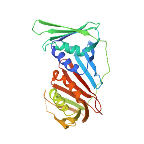
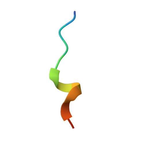
Crystal structure of human PCNA in complex with REV1 PIP-box


Experimental Data Snapshot

  • Method: X-RAY DIFFRACTION
  • Resolution: 2.70 Å
  • R-Value Free: 
    0.261 (Depositor), 0.260 (DCC) 
  • R-Value Work: 
    0.214 (Depositor), 0.214 (DCC) 
  • R-Value Observed: 
    0.216 (Depositor) 

Starting Model: experimental
View more details

wwPDB Validation 3D Report Full Report

Validation slider image for 9VGW

This is version 1.1 of the entry. See complete history

Literature

Identification of a PCNA-binding motif in human translesion DNA polymerase REV1 and structural basis of its interaction with PCNA.

Hishiki, A.Hoshino, N.Okawara, K.Fuchigami, S.Hara, K.Hashimoto, H.

(2025) J Biochem 178: 315-324

  • DOI: https://doi.org/10.1093/jb/mvaf054
  • Primary Citation Related Structures: 
    9VGW

  • PubMed Abstract: 

    REV1 is a eukaryotic error-prone DNA polymerase belonging to the Y-family, with a central role in translesion DNA synthesis (TLS) to continue DNA replication even in the presence of DNA damage in the template strand. TLS is stimulated by monoubiquitination of proliferating cell nuclear antigen (PCNA), a toroidal-shaped protein functioning as a scaffold for DNA polymerases and repair enzymes. Mammals possess four types of Y-family DNA polymerases: Pol η, Pol κ, Pol ι and REV1. Among those, Pol η, Pol κ and Pol ι interact with PCNA through PCNA-binding motifs, low-affinity variants of PCNA-interacting protein box (PIP-box). To date, several studies have reported that REV1 interacts with PCNA, but identified PCNA-binding regions are inconsistent; therefore, a structural basis for interaction between REV1 and PCNA also remains unclear. Here, we identified a signature sequence conserved within vertebrates REV1 responsible for PCNA-binding. Furthermore, we unveiled a mechanism underlying the physical interaction between the PCNA-binding motif of human REV1 and PCNA by X-ray crystallography, thus revealing that REV1 binds to PCNA through a PIP-box variant located in the C-terminal side of the little finger domain. Our study provides a convincing answer for a long-standing controversy regarding the physical interaction between REV1 and PCNA.


  • Organizational Affiliation
    • School of Pharmaceutical Sciences, University of Shizuoka, 52-1 Yada, Suruga-ku, Shizuoka 422-8002, Japan.

Macromolecule Content 

  • Total Structure Weight: 90.93 kDa 
  • Atom Count: 5,940 
  • Modeled Residue Count: 767 
  • Deposited Residue Count: 822 
  • Unique protein chains: 2

Macromolecules

Find similar proteins by:|  3D Structure
Entity ID: 1
MoleculeChains  Sequence LengthOrganismDetailsImage
Proliferating cell nuclear antigen
A, B, C
261Homo sapiensMutation(s): 0 
Gene Names: PCNA
UniProt & NIH Common Fund Data Resources
Find proteins for P12004 (Homo sapiens)
Explore P12004 
Go to UniProtKB:  P12004
PHAROS:  P12004
GTEx:  ENSG00000132646 
Entity Groups
Sequence Clusters30% Identity50% Identity70% Identity90% Identity95% Identity100% Identity
UniProt GroupP12004
Sequence Annotations
Expand
Reference Sequence
Find similar proteins by:  Sequence   |   3D Structure  
Entity ID: 2
MoleculeChains  Sequence LengthOrganismDetailsImage
peptide from DNA repair protein REV1D [auth X],
E [auth Y],
F [auth Z]
13Homo sapiensMutation(s): 0 
EC: 2.7.7
UniProt & NIH Common Fund Data Resources
Find proteins for Q9UBZ9 (Homo sapiens)
Explore Q9UBZ9 
Go to UniProtKB:  Q9UBZ9
PHAROS:  Q9UBZ9
GTEx:  ENSG00000135945 
Entity Groups
Sequence Clusters30% Identity50% Identity70% Identity90% Identity95% Identity100% Identity
UniProt GroupQ9UBZ9
Sequence Annotations
Expand
Reference Sequence

Experimental Data & Validation

Experimental Data

  • Method: X-RAY DIFFRACTION
  • Resolution: 2.70 Å
  • R-Value Free:  0.261 (Depositor), 0.260 (DCC) 
  • R-Value Work:  0.214 (Depositor), 0.214 (DCC) 
  • R-Value Observed: 0.216 (Depositor) 
Space Group: P 21 21 2
Unit Cell:
Length ( Å )Angle ( ˚ )
a = 145.221α = 90
b = 72.508β = 90
c = 83.599γ = 90
Software Package:
Software NamePurpose
REFMACrefinement
XDSdata reduction
SCALAdata scaling
PHASERphasing

Structure Validation

View Full Validation Report


Entry History 

& Funding Information

Deposition Data


Funding OrganizationLocationGrant Number
Japan Society for the Promotion of Science (JSPS)Japan17H06014
Japan Society for the Promotion of Science (JSPS)Japan23K05669

Revision History  (Full details and data files)

  • Version 1.0: 2025-09-10
    Type: Initial release
  • Version 1.1: 2025-11-19
    Changes: Database references