Speciation and structural transformation of a V V -malate complex in the absence and in the presence of a protein: from a dinuclear species to decavanadate.
Paolillo, M., Ferraro, G., Gumerova, N.I., Pisanu, F., Garribba, E., Rompel, A., Merlino, A.(2025) Inorg Chem Front 12: 6503-6518
- PubMed: 40757088 Search on PubMedSearch on PubMed Central
- DOI: https://doi.org/10.1039/d5qi01384d
- Primary Citation Related Structures:
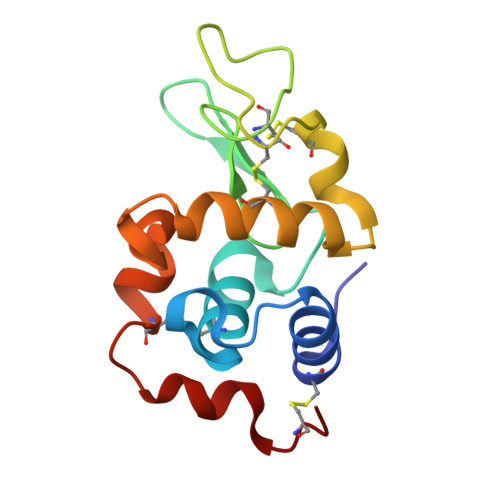
9RBG, 9RBT, 9RBV - PubMed Abstract:
A strategy for the development of new vanadium-based drugs is the preparation of complexes that target proteins and bear molecules involved in the cellular metabolism as ligands, like α-hydroxycarboxylic acids. Based on these premises, this study explores the solution behaviour of the dioxidovanadium(V) complex of malic acid, Cs 2 [V V 2 O 4 (mal) 2 ]·2H 2 O, and its interaction with the model protein lysozyme (HEWL) at room and at physiological temperature using 51 V nuclear magnetic resonance (NMR), electrospray ionisation-mass spectrometry (ESI-MS) and X-ray crystallography. The results show the coexistence in aqueous solution of various molecular species containing two or ten V V centres. In solution these species are formed regardless of the presence of HEWL, while at 37 °C the formation of [V V 10 O 28 ] 6- (V 10 ) is precluded when the protein is present. Crystallographic data reveal that, when protein crystals are incubated with the V compound at room temperature (25 °C) and at pH 4.0, [V IV O] 2+ , [V V 2 O 5 (mal)] 2- , [V V 10 O 26 ] 2- and [V V 10 O 28 ] 6- are bound to the protein, while at 37 °C, under the same conditions, only [V IV O] 2+ interacts with HEWL. [V V 10 O 28 ] 6- can bind the protein both covalently (as [V V 10 O 26 ] 2- ion) and non-covalently. Whereas the transformation of [V V 2 O 4 (mal) 2 ] 2- to [V V 2 O 5 (mal)] 2- is expected on the basis of thermodynamic considerations, the formation of V 10 and of the V 10 -HEWL adduct is not easily predictable. Docking calculations confirm the experimental results and highlight the role of protein-protein interaction in the stabilization of the revealed adduct. This study demonstrates that vanadium compounds can undergo transformation in solution, giving rise to species that interact with proteins through several binding modes and stabilization mechanisms.
- Department of Chemical Sciences, University of Naples Federico II Complesso Universitario di Monte Sant'Angelo Via Cintia I-80126 Napoli Italy antonello.merlino@unina.it.
Organizational Affiliation: