9QEV | pdb_00009qev

A native carbonic anhydrase from Caloramator australiacus


Experimental Data Snapshot

  • Method: X-RAY DIFFRACTION
  • Resolution: 1.11 Å
  • R-Value Free: 
    0.188 (Depositor), 0.191 (DCC) 
  • R-Value Work: 
    0.166 (Depositor), 0.171 (DCC) 
  • R-Value Observed: 
    0.167 (Depositor) 

Starting Model: experimental
View more details

wwPDB Validation 3D Report Full Report

Validation slider image for 9QEV

This is version 1.0 of the entry. See complete history

Literature

Structural and functional characterization of a metagenomically derived gamma-type carbonic anhydrase and its engineering into a hyperthermostable esterase.

Bodourian, C.S.Imran, M.Georgakis, N.D.Papageorgiou, A.C.Labrou, N.E.

(2025) Protein Sci 34: e70396-e70396

  • DOI: https://doi.org/10.1002/pro.70396
  • Primary Citation Related Structures: 
    9QEV, 9QEZ

  • PubMed Abstract: 

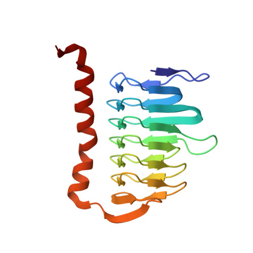
    The 16S microbial community profiling of a metagenomics library from geothermal spring at Lisvori (Lesvos island, Greece) enabled the identification of a putative sequence exhibiting 95% identity to the γ-type carbonic anhydrase (γ-CA) from Caloramator australicus (γ-CaCA). The sequence of γ-CaCA was amplified by PCR, cloned, and expressed in E. coli. Activity assays showed that γ-CaCA possesses very low, but detectable, anhydrase activity, while exhibiting no measurable esterase activity. Differential scanning fluorimetry (DSF) revealed that the enzyme shows high thermal stability with a melting temperature (T m ) approximately 65-75°C in the pH range between 5.5 and 9.0. The structure of γ-CaCA was determined by X-ray crystallography at 1.11 Å resolution, the highest resolution reported so far for a γ-CA. The enzyme was crystallized as a trimer in the crystallographic asymmetric unit and contains three zinc-binding sites, one at each interface of neighboring subunits of the trimer. Structure-based rational design enabled the design and creation of a mutant enzyme (γ-CaCAmut) which possessed a heptapeptide insertion at the active-site loop and two-point mutations. Kinetic analysis demonstrated that γ-CaCAmut was successfully converted into a catalytically active esterase indicating successful activity gain through structure-guided engineering. The thermostability of γ-CaCAmut was significantly increased, aligning with the thermostability typically observed in hyperthermostable enzymes. X-ray crystallographic analysis of the γ-CaCAmut structure at 2.1 Å resolution, provided detailed structural insights into how the mutations impact the overall enzyme structure, function, and thermostability. These findings provide valuable structural and functional insights into γ-CAs and demonstrate a strategy for converting an inactive enzyme into a catalytically active form through rational design.


  • Organizational Affiliation
    • Laboratory of Enzyme Technology, Department of Biotechnology, School of Applied Biology and Biotechnology, Agricultural University of Athens, Athens, Greece.

Macromolecule Content 

  • Total Structure Weight: 58.28 kDa 
  • Atom Count: 4,562 
  • Modeled Residue Count: 507 
  • Deposited Residue Count: 519 
  • Unique protein chains: 1

Macromolecules

Find similar proteins by:|  3D Structure
Entity ID: 1
MoleculeChains  Sequence LengthOrganismDetailsImage
Carbonic anhydrase, family 3
A, B, C
173Caloramator australicusMutation(s): 0 
Gene Names: CAAU_0066
UniProt
Find proteins for G0V3M6 (Caloramator australicus RC3)
Explore G0V3M6 
Go to UniProtKB:  G0V3M6
Entity Groups
Sequence Clusters30% Identity50% Identity70% Identity90% Identity95% Identity100% Identity
UniProt GroupG0V3M6
Sequence Annotations
Expand
Reference Sequence

Small Molecules

Ligands 3 Unique
IDChains Name / Formula / InChI Key2D Diagram3D Interactions
ZN
(Subject of Investigation/LOI)

Query on ZN


Download:Ideal Coordinates CCD File
D [auth A],
L [auth B],
P [auth C]
ZINC ION
Zn
PTFCDOFLOPIGGS-UHFFFAOYSA-N
FMT

Query on FMT


Download:Ideal Coordinates CCD File
K [auth A],
T [auth C]
FORMIC ACID
C H2 O2
BDAGIHXWWSANSR-UHFFFAOYSA-N
NA

Query on NA


Download:Ideal Coordinates CCD File
E [auth A]
F [auth A]
G [auth A]
H [auth A]
I [auth A]
E [auth A],
F [auth A],
G [auth A],
H [auth A],
I [auth A],
J [auth A],
M [auth B],
N [auth B],
O [auth B],
Q [auth C],
R [auth C],
S [auth C]
SODIUM ION
Na
FKNQFGJONOIPTF-UHFFFAOYSA-N

Experimental Data & Validation

Experimental Data

  • Method: X-RAY DIFFRACTION
  • Resolution: 1.11 Å
  • R-Value Free:  0.188 (Depositor), 0.191 (DCC) 
  • R-Value Work:  0.166 (Depositor), 0.171 (DCC) 
  • R-Value Observed: 0.167 (Depositor) 
Space Group: P 21 21 21
Unit Cell:
Length ( Å )Angle ( ˚ )
a = 57.024α = 90
b = 82.847β = 90
c = 98.508γ = 90
Software Package:
Software NamePurpose
PHENIXrefinement
XDSdata reduction
Aimlessdata scaling
PHASERphasing

Structure Validation

View Full Validation Report


Entry History 

& Funding Information

Deposition Data


Funding OrganizationLocationGrant Number
Finnish Cultural FoundationFinland--
iNEXT-DiscoveryEuropean Union--

Revision History  (Full details and data files)

  • Version 1.0: 2025-12-10
    Type: Initial release