


GspB Siglec domain bound to sialyl T antigen linked to serine-FMOC
Morrison, K.M., Martin, K.A., Iverson, T.M.To be published.
Experimental Data Snapshot
Starting Model: experimental
View more details
Entity ID: 1 | |||||
|---|---|---|---|---|---|
| Molecule | Chains | Sequence Length | Organism | Details | Image |
| Platelet binding protein GspB | 127 | Streptococcus | Mutation(s): 0 Gene Names: gspB | 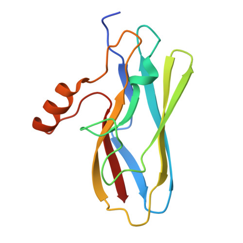 | |
UniProt | |||||
Entity Groups | |||||
| Sequence Clusters | 30% Identity50% Identity70% Identity90% Identity95% Identity100% Identity | ||||
| UniProt Group | A8AWU7 | ||||
Sequence AnnotationsExpand | |||||
Reference Sequence | |||||
| Ligands 4 Unique | |||||
|---|---|---|---|---|---|
| ID | Chains | Name / Formula / InChI Key | 2D Diagram | 3D Interactions | |
| VP1 (Subject of Investigation/LOI) Download:Ideal Coordinates CCD File | G [auth A], J [auth B] | Fluorenylmethyloxycarbonyl chloride C15 H11 Cl O2 IRXSLJNXXZKURP-UHFFFAOYSA-N |  | ||
| SER (Subject of Investigation/LOI) Download:Ideal Coordinates CCD File | H [auth A], K [auth B] | SERINE C3 H7 N O3 MTCFGRXMJLQNBG-REOHCLBHSA-N |  | ||
| GOL (Subject of Investigation/LOI) Download:Ideal Coordinates CCD File | E [auth A], F [auth A] | GLYCEROL C3 H8 O3 PEDCQBHIVMGVHV-UHFFFAOYSA-N |  | ||
| CA Download:Ideal Coordinates CCD File | I [auth A], L [auth B] | CALCIUM ION Ca BHPQYMZQTOCNFJ-UHFFFAOYSA-N |  | ||
| Length ( Å ) | Angle ( ˚ ) |
|---|---|
| a = 37.987 | α = 70.2 |
| b = 38.111 | β = 68.01 |
| c = 47.054 | γ = 62.08 |
| Software Name | Purpose |
|---|---|
| PHENIX | refinement |
| HKL-2000 | data reduction |
| HKL-2000 | data scaling |
| PHASER | phasing |
| Funding Organization | Location | Grant Number |
|---|---|---|
| National Institutes of Health/National Institute of General Medical Sciences (NIH/NIGMS) | United States | -- |


