9PQV | pdb_00009pqv

Crystal structure of C. elegans PUF-3 in complex with RNA II-2


Experimental Data Snapshot

  • Method: X-RAY DIFFRACTION
  • Resolution: 2.92 Å
  • R-Value Free: 
    0.214 (Depositor), 0.211 (DCC) 
  • R-Value Work: 
    0.196 (Depositor), 0.195 (DCC) 
  • R-Value Observed: 
    0.197 (Depositor) 

Starting Model: experimental
View more details

wwPDB Validation   3D Report Full Report


Ligand Structure Quality Assessment 


This is version 1.1 of the entry. See complete history


Literature

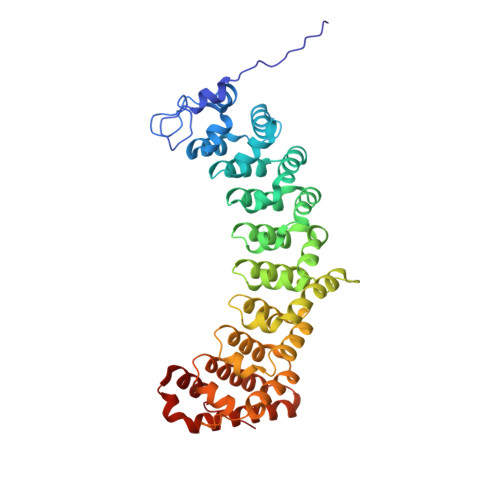
Crystal structures of Caenorhabditis elegans PUF-3 depict plasticity of RNA recognition that enables germline gene regulation.

Zhang, Y.Kennedy, F.D.Hall, T.M.T.

(2026) Nucleic Acids Res 54

  • DOI: https://doi.org/10.1093/nar/gkaf1431
  • Primary Citation of Related Structures:  
    9PMM, 9PQV, 9PQW, 9PSA, 9PSB, 9PSC, 9PTD, 9PTG, 9PTJ

  • PubMed Abstract: 

    Prototypical PUF proteins are known for modular, sequence-specific RNA recognition. A single PUF protein in Drosophila melanogaster Pumilio and two closely related proteins in mammals, PUM1 and PUM2, are representative. In contrast, Caenorhabditis elegans PUF proteins have evolved to include four subfamilies of single-stranded RNA-binding proteins that recognize distinct sequence elements. Here, we provide an in-depth structural and biochemical analysis of C. elegans PUF-3, a member of the subfamily lacking structural information. We determined crystal structures of PUF-3 in complex with RNAs representing three classes of binding elements. These structures and quantitative RNA-binding assays fine-tune the features of PUF-3 RNA recognition and demonstrate how PUF-3 binds to varied target RNA elements by accommodating divergent sequences between conserved features. Our analyses here and published biological data suggest that PUF-3 may redundantly regulate RNA targets with other PUF proteins. We determined the in vitro binding affinities of C. elegans PUF protein subfamily representatives for the range of corresponding RNA elements. We find that PUF-3 and FBF-2 have broader RNA recognition, whereas PUF-8 and PUF-6 are highly selective. These subfamily recognition properties and protein expression patterns support a working model for PUF protein activities in different regions of the C. elegans germline.


  • Organizational Affiliation
    • Epigenetics and RNA Biology Laboratory, National Institute of Environmental Health Sciences, National Institutes of Health, Research Triangle Park, NC 27709, United States.

Macromolecules

Find similar proteins by:  (by identity cutoff)  |  3D Structure
Entity ID: 1
MoleculeChains Sequence LengthOrganismDetailsImage
PUM-HD domain-containing protein411Caenorhabditis elegansMutation(s): 0 
Gene Names: puf-3CELE_Y45F10A.2Y45F10A.2
UniProt
Find proteins for Q9U2G4 (Caenorhabditis elegans)
Explore Q9U2G4 
Go to UniProtKB:  Q9U2G4
Entity Groups  
Sequence Clusters30% Identity50% Identity70% Identity90% Identity95% Identity100% Identity
UniProt GroupQ9U2G4
Sequence Annotations
Expand
  • Reference Sequence

Find similar nucleic acids by:  Sequence   |   3D Structure  

Entity ID: 2
MoleculeChains LengthOrganismImage
RNA II-210Caenorhabditis elegans
Sequence Annotations
Expand
  • Reference Sequence
Small Molecules
Ligands 2 Unique
IDChains Name / Formula / InChI Key2D Diagram3D Interactions
GOL (Subject of Investigation/LOI)
Query on GOL

Download Ideal Coordinates CCD File 
C [auth A],
I [auth A],
L [auth A],
N [auth A]
GLYCEROL
C3 H8 O3
PEDCQBHIVMGVHV-UHFFFAOYSA-N
FMT (Subject of Investigation/LOI)
Query on FMT

Download Ideal Coordinates CCD File 
D [auth A]
E [auth A]
F [auth A]
G [auth A]
H [auth A]
D [auth A],
E [auth A],
F [auth A],
G [auth A],
H [auth A],
J [auth A],
K [auth A],
M [auth A],
O [auth A],
P [auth A],
Q [auth A],
R [auth B],
S [auth B]
FORMIC ACID
C H2 O2
BDAGIHXWWSANSR-UHFFFAOYSA-N
Experimental Data & Validation

Experimental Data

  • Method: X-RAY DIFFRACTION
  • Resolution: 2.92 Å
  • R-Value Free:  0.214 (Depositor), 0.211 (DCC) 
  • R-Value Work:  0.196 (Depositor), 0.195 (DCC) 
  • R-Value Observed: 0.197 (Depositor) 
Space Group: P 32 2 1
Unit Cell:
Length ( Å )Angle ( ˚ )
a = 161.758α = 90
b = 161.758β = 90
c = 100.259γ = 120
Software Package:
Software NamePurpose
PHENIXrefinement
Aimlessdata scaling
PDB_EXTRACTdata extraction
XDSdata reduction
PHASERphasing

Structure Validation

View Full Validation Report


Ligand Structure Quality Assessment 


Entry History & Funding Information

Deposition Data


Funding OrganizationLocationGrant Number
National Institutes of Health/National Institute of Environmental Health Sciences (NIH/NIEHS)United States1ZIA-ES050165

Revision History  (Full details and data files)

  • Version 1.0: 2025-12-17
    Type: Initial release
  • Version 1.1: 2026-01-28
    Changes: Database references