9PHA | pdb_00009pha

CryoEM structure of the YonE portal protein from Bacillus phage SPbeta


Experimental Data Snapshot

  • Method: ELECTRON MICROSCOPY
  • Resolution: 3.30 Å
  • Aggregation State: PARTICLE 
  • Reconstruction Method: SINGLE PARTICLE 

wwPDB Validation 3D Report Full Report

Validation slider image for 9PHA

This is version 1.2 of the entry. See complete history

Literature

Molecular characterisation of the Bacillus subtilis SpbK antiphage defence system.

Mishra, B.P.Loyo, C.L.Cai, Y.Litfin, T.Miraj, G.Brillault, L.Masic, V.Mosaiab, T.Rajaratnam, P.Rudrawar, S.Gu, W.Kobe, B.Gerdt, J.P.Grossman, A.D.Shi, Y.Ve, T.

(2025) Nat Commun 17: 1051-1051

  • DOI: https://doi.org/10.1038/s41467-025-67810-5
  • Primary Citation Related Structures: 
    9PHA, 9PHB

  • PubMed Abstract: 

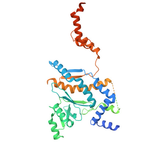
    Bacteria have a variety of mechanisms for limiting predation by phages. SpbK is a Toll/interleukin-1 receptor (TIR) domain-containing antiphage defence protein from Bacillus subtilis that provides protection against the temperate phage SPβ via abortive infection. Here we structurally characterise SpbK and its interaction with the SPβ protein YonE. We demonstrate that SpbK is an NADase that produces both ADP-ribose (ADPR) and canonical cyclic ADPR with a N1-glycosidic bond (cADPR, also referred to as N1-cADPR). Combining cryo-EM, in silico predictions, site-directed mutagenesis, and phage infection assays, we show that formation of two-stranded head-to-tail assemblies of SpbK TIR domains is required for both NADase activity and antiphage defence. We also demonstrate that YonE is a dodecameric portal protein that activates the NADase function of SpbK by facilitating TIR domain clustering. Collectively, our results provide insight into how bacterial TIR NADases recognise phage infection.


  • Organizational Affiliation
    • Institute for Biomedicine and Glycomics, Griffith University, Gold Coast, QLD, Australia.

Macromolecule Content 

  • Total Structure Weight: 666.34 kDa 
  • Atom Count: 30,144 
  • Modeled Residue Count: 3,600 
  • Deposited Residue Count: 5,820 
  • Unique protein chains: 1

Macromolecules

Find similar proteins by:|  3D Structure
Entity ID: 1
MoleculeChains  Sequence LengthOrganismDetailsImage
SPbeta prophage-derived uncharacterized protein YonE
A, B, C, D, E
A, B, C, D, E, F, G, H, I, J, K, L
485Spbetavirus SPbetaMutation(s): 0 
Gene Names: yonEBSU21120
UniProt
Find proteins for O31953 (Bacillus subtilis (strain 168))
Explore O31953 
Go to UniProtKB:  O31953
Entity Groups
Sequence Clusters30% Identity50% Identity70% Identity90% Identity95% Identity100% Identity
UniProt GroupO31953
Sequence Annotations
Expand
Reference Sequence

Experimental Data & Validation

Experimental Data

  • Method: ELECTRON MICROSCOPY
  • Resolution: 3.30 Å
  • Aggregation State: PARTICLE 
  • Reconstruction Method: SINGLE PARTICLE 
EM Software:
TaskSoftware PackageVersion
MODEL REFINEMENTPHENIX1.20.1_4487

Structure Validation

View Full Validation Report


Entry History 

& Funding Information

Deposition Data


Funding OrganizationLocationGrant Number
Australian Research Council (ARC)AustraliaFT200100572
National Health and Medical Research Council (NHMRC, Australia)Australia1196590

Revision History  (Full details and data files)

  • Version 1.0: 2025-12-31
    Type: Initial release
  • Version 1.1: 2026-01-14
    Changes: Data collection, Database references
  • Version 1.2: 2026-02-04
    Changes: Data collection, Database references