9P97 | pdb_00009p97

CryoEM structure of the closed integrin alphaEbeta7 bound to fab LF61


Experimental Data Snapshot

  • Method: ELECTRON MICROSCOPY
  • Resolution: 2.92 Å
  • Aggregation State: PARTICLE 
  • Reconstruction Method: SINGLE PARTICLE 

wwPDB Validation   3D Report Full Report


This is version 1.0 of the entry. See complete history


Literature

Molecular exaptation by the integrin alpha I domain.

Hollis, J.A.Chan, M.C.Malik, H.S.Campbell, M.G.

(2025) Sci Adv 11: eadx9567-eadx9567

  • DOI: https://doi.org/10.1126/sciadv.adx9567
  • Primary Citation of Related Structures:  
    9P95, 9P96, 9P97, 9P98, 9P99

  • PubMed Abstract: 

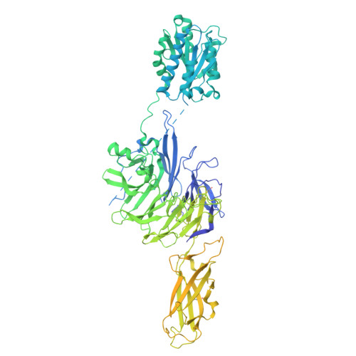
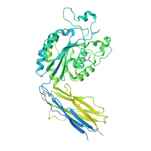
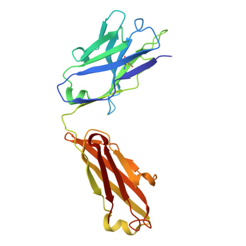
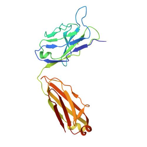
    Integrins bind ligands between their alpha (α) and beta (β) subunits and transmit signals through conformational changes. Early in chordate evolution, some α subunits acquired an "inserted" (I) domain that expanded integrin's ligand-binding repertoire but obstructed the ancestral ligand pocket, seemingly blocking conventional integrin activation. Here, we compare cryo-electron microscopy structures of apo and ligand-bound states of the I domain-containing αEβ 7 integrin and the I domain-lacking α 4 β 7 integrin to illuminate how the I domain intrinsically mimics an extrinsic ligand to preserve integrin function. We trace the I domain's evolutionary origin to an ancestral collagen-collagen interaction domain, identifying an ancient molecular exaptation that facilitated integrin activation immediately upon I domain insertion. Our analyses reveal the evolutionary and biochemical basis of expanded cellular communication in vertebrates.


  • Organizational Affiliation
    • Division of Basic Sciences, Fred Hutchinson Cancer Center, Seattle, WA 98109, USA.

Macromolecules
Find similar proteins by:  (by identity cutoff)  |  3D Structure
Entity ID: 1
MoleculeChains Sequence LengthOrganismDetailsImage
Integrin alpha-E1,176Homo sapiensMutation(s): 0 
Gene Names: ITGAE
UniProt & NIH Common Fund Data Resources
Find proteins for P38570 (Homo sapiens)
Explore P38570 
Go to UniProtKB:  P38570
PHAROS:  P38570
GTEx:  ENSG00000083457 
Entity Groups  
Sequence Clusters30% Identity50% Identity70% Identity90% Identity95% Identity100% Identity
UniProt GroupP38570
Glycosylation
Glycosylation Sites: 5Go to GlyGen: P38570-1
Sequence Annotations
Expand
  • Reference Sequence
Find similar proteins by:  (by identity cutoff)  |  3D Structure
Entity ID: 2
MoleculeChains Sequence LengthOrganismDetailsImage
Integrin beta-7776Homo sapiensMutation(s): 0 
Gene Names: ITGB7
UniProt & NIH Common Fund Data Resources
Find proteins for P26010 (Homo sapiens)
Explore P26010 
Go to UniProtKB:  P26010
PHAROS:  P26010
GTEx:  ENSG00000139626 
Entity Groups  
Sequence Clusters30% Identity50% Identity70% Identity90% Identity95% Identity100% Identity
UniProt GroupP26010
Glycosylation
Glycosylation Sites: 2Go to GlyGen: P26010-1
Sequence Annotations
Expand
  • Reference Sequence
Find similar proteins by:  (by identity cutoff)  |  3D Structure
Entity ID: 3
MoleculeChains Sequence LengthOrganismDetailsImage
Fab LF61 Heavy ChainC [auth H]220Mus musculusMutation(s): 0 
Entity Groups  
Sequence Clusters30% Identity50% Identity70% Identity90% Identity95% Identity100% Identity
Sequence Annotations
Expand
  • Reference Sequence
Find similar proteins by:  (by identity cutoff)  |  3D Structure
Entity ID: 4
MoleculeChains Sequence LengthOrganismDetailsImage
Fab LF61 Light ChainD [auth L]215Mus musculusMutation(s): 0 
Entity Groups  
Sequence Clusters30% Identity50% Identity70% Identity90% Identity95% Identity100% Identity
Sequence Annotations
Expand
  • Reference Sequence
Oligosaccharides

Help

Entity ID: 5
MoleculeChains Length2D Diagram Glycosylation3D Interactions
alpha-D-mannopyranose-(1-3)-[alpha-D-mannopyranose-(1-6)]beta-D-mannopyranose-(1-4)-2-acetamido-2-deoxy-beta-D-glucopyranose-(1-4)-2-acetamido-2-deoxy-beta-D-glucopyranoseE [auth C]5N-Glycosylation
Glycosylation Resources
GlyTouCan:  G22768VO
GlyCosmos:  G22768VO
GlyGen:  G22768VO
Entity ID: 6
MoleculeChains Length2D Diagram Glycosylation3D Interactions
2-acetamido-2-deoxy-beta-D-glucopyranose-(1-4)-2-acetamido-2-deoxy-beta-D-glucopyranoseF [auth D],
H [auth F]
2N-Glycosylation
Glycosylation Resources
GlyTouCan:  G42666HT
GlyCosmos:  G42666HT
GlyGen:  G42666HT
Entity ID: 7
MoleculeChains Length2D Diagram Glycosylation3D Interactions
beta-D-mannopyranose-(1-4)-2-acetamido-2-deoxy-beta-D-glucopyranose-(1-4)-2-acetamido-2-deoxy-beta-D-glucopyranoseG [auth E]3N-Glycosylation
Glycosylation Resources
GlyTouCan:  G15407YE
GlyCosmos:  G15407YE
GlyGen:  G15407YE
Small Molecules
Ligands 2 Unique
IDChains Name / Formula / InChI Key2D Diagram3D Interactions
NAG
Query on NAG

Download Ideal Coordinates CCD File 
I [auth A],
J [auth A],
P [auth B]
2-acetamido-2-deoxy-beta-D-glucopyranose
C8 H15 N O6
OVRNDRQMDRJTHS-FMDGEEDCSA-N
CA
Query on CA

Download Ideal Coordinates CCD File 
K [auth A]
L [auth A]
M [auth A]
N [auth B]
O [auth B]
K [auth A],
L [auth A],
M [auth A],
N [auth B],
O [auth B],
Q [auth B]
CALCIUM ION
Ca
BHPQYMZQTOCNFJ-UHFFFAOYSA-N
Experimental Data & Validation

Experimental Data

  • Method: ELECTRON MICROSCOPY
  • Resolution: 2.92 Å
  • Aggregation State: PARTICLE 
  • Reconstruction Method: SINGLE PARTICLE 
EM Software:
TaskSoftware PackageVersion
MODEL REFINEMENTPHENIX1.20.1_4487
RECONSTRUCTIONcryoSPARC

Structure Validation

View Full Validation Report


Entry History & Funding Information

Deposition Data


Funding OrganizationLocationGrant Number
National Institutes of Health/National Institute of General Medical Sciences (NIH/NIGMS)United StatesR35GM147414
National Science Foundation (NSF, United States)United States--
National Institutes of Health/National Institute of General Medical Sciences (NIH/NIGMS)United StatesT32GM008268

Revision History  (Full details and data files)

  • Version 1.0: 2025-09-24
    Type: Initial release