9P0X | pdb_00009p0x

Nanodisc-embedded human TF/FVIIa/XK1 in complex with 10H10 Fab (nanodisc-subtracted)


Experimental Data Snapshot

  • Method: ELECTRON MICROSCOPY
  • Resolution: 3.70 Å
  • Aggregation State: PARTICLE 
  • Reconstruction Method: SINGLE PARTICLE 

Starting Model: experimental
View more details

wwPDB Validation   3D Report Full Report


This is version 1.1 of the entry. See complete history


Literature

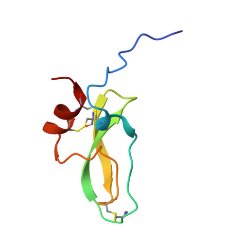
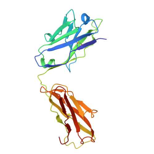
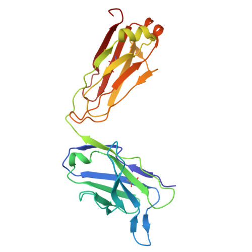
Cryo-EM structure of the tissue factor/factor VIIa complex with a factor X mimetic reveals a novel allosteric mechanism.

Sedzro, J.C.Photenhauer, A.L.Birkle, F.Meze, K.Mortenson, A.Duckworth, C.Wen, P.C.Kearns, S.Cianfrocco, M.A.Tajkhorshid, E.Ohi, M.D.Morrissey, J.H.

(2025) Blood 146: 2833-2842

  • DOI: https://doi.org/10.1182/blood.2025029430
  • Primary Citation of Related Structures:  
    9P0X

  • PubMed Abstract: 

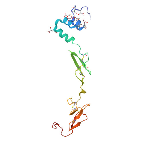
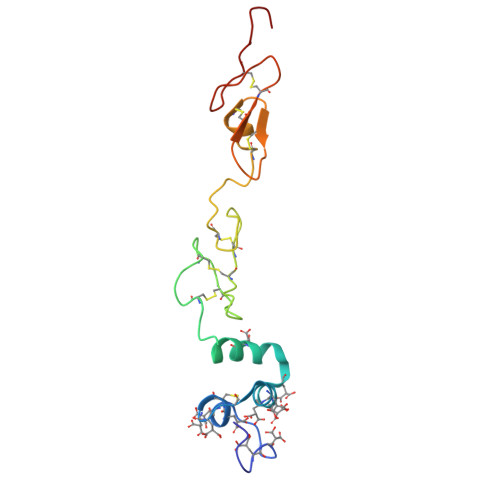
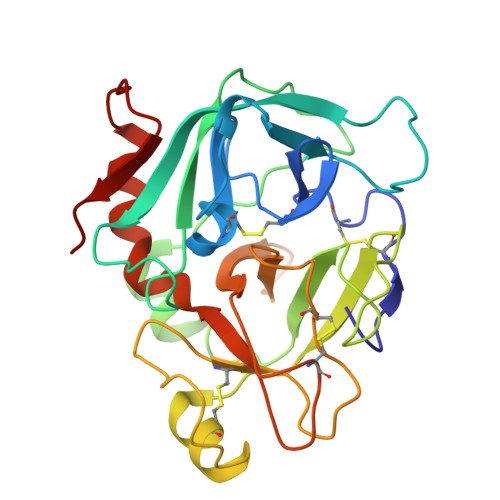
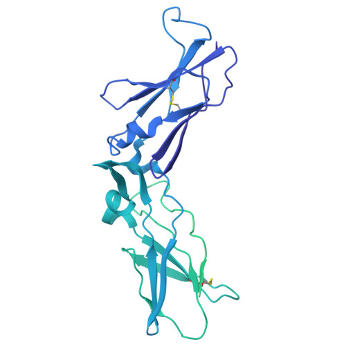
    Blood clotting is triggered in hemostasis and thrombosis when the membrane-bound tissue factor (TF)/factor VIIa (FVIIa) complex activates factor X (FX). There are no structures of TF/FVIIa on membranes, with or without FX. Using cryo-EM to address this gap, we assembled TF/FVIIa complexes on nanoscale membrane bilayers (nanodiscs), bound to XK1 and an antibody fragment. XK1 is a FX mimetic whose protease domain is replaced by the first Kunitz-type (K1) domain of tissue factor pathway inhibitor, while 10H10 is a non-inhibitory, anti-TF antibody. We determined a cryo-EM structure of a TF/FVIIa/XK1/10H10/nanodisc complex with a resolution of 3.7 Å, allowing us to model all the protein backbones. TF/FVIIa extends perpendicularly from the membrane, interacting with a "handle shaped" XK1 at two locations: the K1 domain docks into FVIIa's active site, while the γ-carboxyglutamate-rich (GLA) domain binds to TF's substrate-binding exosite. The FX and FVIIa GLA domains also contact each other and the membrane surface. Except for a minor contact between the first epidermal growth factor (EGF)-like domain of XK1 and TF, the rest of the FX light chain does not interact with TF/FVIIa. The structure reveals a previously unrecognized, membrane-dependent allosteric activation mechanism between FVIIa and TF where a serine-rich loop in TF that partially obscures the TF exosite must undergo a shift to allow access of the FX GLA domain to its final binding location on the membrane-bound TF/FVIIa complex. This mechanism also provides a novel explanation for the otherwise puzzling phenomenon of TF encryption/decryption on cell surfaces.


  • Organizational Affiliation
    • University of Michigan, Ann Arbor, Michigan, United States.

Macromolecules
Find similar proteins by:  (by identity cutoff)  |  3D Structure
Entity ID: 1
MoleculeChains Sequence LengthOrganismDetailsImage
Factor VII light chainA [auth V]142Homo sapiensMutation(s): 0 
Gene Names: F7
EC: 3.4.21.21
UniProt & NIH Common Fund Data Resources
Find proteins for P08709 (Homo sapiens)
Explore P08709 
Go to UniProtKB:  P08709
PHAROS:  P08709
GTEx:  ENSG00000057593 
Entity Groups  
Sequence Clusters30% Identity50% Identity70% Identity90% Identity95% Identity100% Identity
UniProt GroupP08709
Glycosylation
Glycosylation Sites: 2Go to GlyGen: P08709-1
Sequence Annotations
Expand
  • Reference Sequence
Find similar proteins by:  (by identity cutoff)  |  3D Structure
Entity ID: 2
MoleculeChains Sequence LengthOrganismDetailsImage
Factor X light chainB [auth X]131Homo sapiensMutation(s): 0 
Gene Names: F10
EC: 3.4.21.6
UniProt & NIH Common Fund Data Resources
Find proteins for P00742 (Homo sapiens)
Explore P00742 
Go to UniProtKB:  P00742
PHAROS:  P00742
GTEx:  ENSG00000126218 
Entity Groups  
Sequence Clusters30% Identity50% Identity70% Identity90% Identity95% Identity100% Identity
UniProt GroupP00742
Sequence Annotations
Expand
  • Reference Sequence
Find similar proteins by:  (by identity cutoff)  |  3D Structure
Entity ID: 3
MoleculeChains Sequence LengthOrganismDetailsImage
Coagulation factor VII Heavy ChainC [auth S]254Homo sapiensMutation(s): 0 
Gene Names: F7
EC: 3.4.21.21
UniProt & NIH Common Fund Data Resources
Find proteins for P08709 (Homo sapiens)
Explore P08709 
Go to UniProtKB:  P08709
PHAROS:  P08709
GTEx:  ENSG00000057593 
Entity Groups  
Sequence Clusters30% Identity50% Identity70% Identity90% Identity95% Identity100% Identity
UniProt GroupP08709
Sequence Annotations
Expand
  • Reference Sequence
Find similar proteins by:  (by identity cutoff)  |  3D Structure
Entity ID: 4
MoleculeChains Sequence LengthOrganismDetailsImage
Tissue factor,Maltose/maltodextrin-binding periplasmic proteinD [auth T]611Homo sapiensEscherichia coli
This entity is chimeric
Mutation(s): 0 
Gene Names: F3malEZ5632ECs5017
UniProt & NIH Common Fund Data Resources
Find proteins for P0AEX9 (Escherichia coli (strain K12))
Explore P0AEX9 
Go to UniProtKB:  P0AEX9
Find proteins for P13726 (Homo sapiens)
Explore P13726 
Go to UniProtKB:  P13726
PHAROS:  P13726
GTEx:  ENSG00000117525 
Entity Groups  
Sequence Clusters30% Identity50% Identity70% Identity90% Identity95% Identity100% Identity
UniProt GroupsP13726P0AEX9
Sequence Annotations
Expand
  • Reference Sequence
Find similar proteins by:  (by identity cutoff)  |  3D Structure
Entity ID: 5
MoleculeChains Sequence LengthOrganismDetailsImage
Tissue factor pathway inhibitorE [auth K]68Homo sapiensMutation(s): 0 
Gene Names: TFPILACITFPI1
UniProt & NIH Common Fund Data Resources
Find proteins for P10646 (Homo sapiens)
Explore P10646 
Go to UniProtKB:  P10646
PHAROS:  P10646
GTEx:  ENSG00000003436 
Entity Groups  
Sequence Clusters30% Identity50% Identity70% Identity90% Identity95% Identity100% Identity
UniProt GroupP10646
Sequence Annotations
Expand
  • Reference Sequence
Find similar proteins by:  (by identity cutoff)  |  3D Structure
Entity ID: 6
MoleculeChains Sequence LengthOrganismDetailsImage
Human 10H10 antibody Fab heavy chainF [auth H]221Mus musculusMutation(s): 0 
Entity Groups  
Sequence Clusters30% Identity50% Identity70% Identity90% Identity95% Identity100% Identity
Sequence Annotations
Expand
  • Reference Sequence
Find similar proteins by:  (by identity cutoff)  |  3D Structure
Entity ID: 7
MoleculeChains Sequence LengthOrganismDetailsImage
Human 10H10 antibody Fab light chainG [auth L]220Mus musculusMutation(s): 0 
Entity Groups  
Sequence Clusters30% Identity50% Identity70% Identity90% Identity95% Identity100% Identity
Sequence Annotations
Expand
  • Reference Sequence
Small Molecules
Ligands 4 Unique
IDChains Name / Formula / InChI Key2D Diagram3D Interactions
BGC
Query on BGC

Download Ideal Coordinates CCD File 
O [auth V]beta-D-glucopyranose
C6 H12 O6
WQZGKKKJIJFFOK-VFUOTHLCSA-N
FUC
Query on FUC

Download Ideal Coordinates CCD File 
P [auth V]alpha-L-fucopyranose
C6 H12 O5
SHZGCJCMOBCMKK-SXUWKVJYSA-N
CA
Query on CA

Download Ideal Coordinates CCD File 
I [auth V]
J [auth V]
L [auth V]
M [auth V]
Q [auth V]
I [auth V],
J [auth V],
L [auth V],
M [auth V],
Q [auth V],
R [auth X],
S [auth X],
T [auth X],
U [auth X],
V [auth X],
W [auth X],
Y [auth S]
CALCIUM ION
Ca
BHPQYMZQTOCNFJ-UHFFFAOYSA-N
MG
Query on MG

Download Ideal Coordinates CCD File 
H [auth V],
K [auth V],
N [auth V],
X
MAGNESIUM ION
Mg
JLVVSXFLKOJNIY-UHFFFAOYSA-N
Modified Residues  1 Unique
IDChains TypeFormula2D DiagramParent
CGU
Query on CGU
A [auth V]L-PEPTIDE LINKINGC6 H9 N O6GLU
Experimental Data & Validation

Experimental Data

  • Method: ELECTRON MICROSCOPY
  • Resolution: 3.70 Å
  • Aggregation State: PARTICLE 
  • Reconstruction Method: SINGLE PARTICLE 
EM Software:
TaskSoftware PackageVersion
MODEL REFINEMENTPHENIX1.21.2_5419
RECONSTRUCTIONcryoSPARC

Structure Validation

View Full Validation Report


Entry History & Funding Information

Deposition Data


Funding OrganizationLocationGrant Number
National Institutes of Health/National Heart, Lung, and Blood Institute (NIH/NHLBI)United StatesR35 HL135823
National Institutes of Health/National Heart, Lung, and Blood Institute (NIH/NHLBI)United StatesR35 HL171334
National Institutes of Health/Office of the DirectorUnited StatesS10OD030275
National Institutes of Health/National Institute of General Medical Sciences (NIH/NIGMS)United StatesR24 GM145965
American Heart AssociationUnited StatesN028347

Revision History  (Full details and data files)

  • Version 1.0: 2025-08-27
    Type: Initial release
  • Version 1.1: 2025-12-17
    Changes: Data collection, Database references