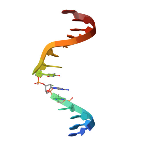
9OSO | pdb_00009oso

The specificity and structure of DNA crosslinking by a gut bacterial genotoxin

  • Classification: DNA
  • Organism(s): Escherichia coli
  • Expression System: Escherichia coli
  • Mutation(s): No 

  • Deposited: 2025-05-25 Released: 2025-11-19 
  • Deposition Author(s): D'Souza, V.D., Balskus, E.P.
  • Funding Organization(s): National Institutes of Health/National Cancer Institute (NIH/NCI), Howard Hughes Medical Institute (HHMI)

Experimental Data Snapshot

  • Method: SOLUTION NMR
  • Conformers Calculated: 50 
  • Conformers Submitted: 10 
  • Selection Criteria: structures with the lowest energy 

wwPDB Validation 3D Report Full Report

Validation slider image for 9OSO

This is version 1.1 of the entry. See complete history

Literature

The specificity and structure of DNA cross-linking by the gut bacterial genotoxin colibactin.

Carlson, E.S.Haslecker, R.Lecchi, C.Aguilar Ramos, M.A.Vennelakanti, V.Honaker, L.Stornetta, A.Millan, E.S.Johnson, B.A.Kulik, H.J.Balbo, S.Villalta, P.W.D'Souza, V.M.Balskus, E.P.

(2025) Science 390: eady3571-eady3571

  • DOI: https://doi.org/10.1126/science.ady3571
  • Primary Citation Related Structures: 
    9OSO

  • PubMed Abstract: 

    Accumulating evidence has connected the chemically unstable, DNA-damaging gut bacterial natural product colibactin to colorectal cancer, including the identification of mutational signatures that are thought to arise from colibactin-DNA interstrand cross-links (ICLs). However, we currently lack direct information regarding the structure of this lesion. In this work, we combined mass spectrometry and nuclear magnetic resonance spectroscopy to elucidate the specificity and structure of the colibactin-DNA ICL. We found that colibactin alkylates within the minor groove of adenine- and thymine-rich DNA, explaining the origins of mutational signatures. Unexpectedly, we discovered that the chemically unstable central motif of colibactin mediates the sequence specificity of cross-linking. By directly elucidating colibactin's interactions with DNA, this work enhances our understanding of the structure and genotoxic mechanisms of this cancer-linked gut bacterial natural product.


  • Organizational Affiliation
    • Department of Chemistry and Chemical Biology, Harvard University, Cambridge, MA, USA.

Macromolecule Content 

  • Total Structure Weight: 9.37 kDa 
  • Atom Count: 624 
  • Modeled Residue Count: 28 
  • Deposited Residue Count: 28 
  • Unique nucleic acid chains: 1

Macromolecules

Find similar nucleic acids by:  Sequence
Entity ID: 1
MoleculeChains LengthOrganismImage
DNA-COLIBACTIN
A, B
14Escherichia coli
Sequence Annotations
Expand
Reference Sequence

Small Molecules

Ligands 1 Unique
IDChains Name / Formula / InChI Key2D Diagram3D Interactions
A1CEC
(Subject of Investigation/LOI)

Query on A1CEC


Download:Ideal Coordinates CCD File
C [auth A]N-({4-[(2R)-2-amino-2-{4-[(2-{(4Z)-2-methyl-4-[(5S)-5-methylpyrrolidin-2-ylidene]-5-oxo-4,5-dihydro-1H-pyrrol-3-yl}acetamido)acetyl]-1,3-thiazol-2-yl}acetyl]-1,3-thiazol-2-yl}methyl)-2-{(4Z)-2-methyl-4-[(5S)-5-methylpyrrolidin-2-ylidene]-5-oxo-4,5-dihydro-1H-pyrrol-3-yl}acetamide
C35 H39 N9 O6 S2
VHOMKILVRMYTBO-ORFCZNMFSA-N

Experimental Data & Validation

Experimental Data

  • Method: SOLUTION NMR
  • Conformers Calculated: 50 
  • Conformers Submitted: 10 
  • Selection Criteria: structures with the lowest energy 

Structure Validation

View Full Validation Report


Entry History 

& Funding Information

Deposition Data


Funding OrganizationLocationGrant Number
National Institutes of Health/National Cancer Institute (NIH/NCI)United StatesR01CA208834
Howard Hughes Medical Institute (HHMI)United States55108516

Revision History  (Full details and data files)

  • Version 1.0: 2025-11-19
    Type: Initial release
  • Version 1.1: 2025-12-17
    Changes: Database references