9NNS | pdb_00009nns

Flavin-dependent N5-ornithine monooxygenase EtcB


Experimental Data Snapshot

  • Method: X-RAY DIFFRACTION
  • Resolution: 3.00 Å
  • R-Value Free: 
    0.350 (Depositor), 0.350 (DCC) 
  • R-Value Work: 
    0.253 (Depositor), 0.253 (DCC) 
  • R-Value Observed: 
    0.257 (Depositor) 

Starting Model: in silico
View more details

wwPDB Validation   3D Report Full Report


Ligand Structure Quality Assessment 


This is version 1.2 of the entry. See complete history


Literature

Redefining the role of the EryM acetyltransferase in natural product biosynthetic pathways.

Li, Y.Liu, X.Harris, N.R.Roberts, J.R.Valdivia, E.M.Ji, X.Smith, J.L.

(2025) Structure 33: 1352-1361.e3

  • DOI: https://doi.org/10.1016/j.str.2025.05.011
  • Primary Citation of Related Structures:  
    9NNQ, 9NNR, 9NNS, 9OA7

  • PubMed Abstract: 

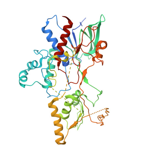
    The GNAT (GCN5-related N-acetyltransferase) superfamily comprises enzymes with a conserved fold and diverse catalytic activities, including primarily acyl transfer, with a few examples of decarboxylation. EryM, a GNAT from Saccharopolyspora erythraea, has been implicated in both erythromycin and erythrochelin biosynthesis, with dual functionality as an acetyltransferase and a decarboxylase. Despite an historical association with malonyl-coenzyme A decarboxylation activity, this dual activity has remained enigmatic as its close homologs were identified with only acyl transfer activity. Here, functional assays demonstrate that EryM catalyzes acyl transfer but lacks decarboxylation activity, challenging long-standing assumptions about its biosynthetic role. Crystal structures of EryM and an acetyl-CoA complex and comparison with homologs in siderophore pathways reveal a conserved catalytic pocket with an essential His and identically positioned side chains common to GNAT enzymes for N-acyl transfer from CoA to primary hydroxylamine substrates. Bioinformatic analysis defines a large GNAT subfamily broadly distributed in the microbial world.


  • Organizational Affiliation
    • Life Sciences Institute, University of Michigan, Ann Arbor, MI 48109, USA; Department of Biophysics, University of Michigan, Ann Arbor, MI 48109, USA.

Macromolecules
Find similar proteins by:  (by identity cutoff)  |  3D Structure
Entity ID: 1
MoleculeChains Sequence LengthOrganismDetailsImage
L-lysine N6-monooxygenase MbtG
A, B
460Saccharopolyspora erythraeaMutation(s): 0 
Gene Names: SACE_3033
EC: 1.14.13.59
UniProt
Find proteins for A4FE36 (Saccharopolyspora erythraea (strain ATCC 11635 / DSM 40517 / JCM 4748 / NBRC 13426 / NCIMB 8594 / NRRL 2338))
Explore A4FE36 
Go to UniProtKB:  A4FE36
Entity Groups  
Sequence Clusters30% Identity50% Identity70% Identity90% Identity95% Identity100% Identity
UniProt GroupA4FE36
Sequence Annotations
Expand
  • Reference Sequence
Experimental Data & Validation

Experimental Data

  • Method: X-RAY DIFFRACTION
  • Resolution: 3.00 Å
  • R-Value Free:  0.350 (Depositor), 0.350 (DCC) 
  • R-Value Work:  0.253 (Depositor), 0.253 (DCC) 
  • R-Value Observed: 0.257 (Depositor) 
Space Group: P 21 2 21
Unit Cell:
Length ( Å )Angle ( ˚ )
a = 76.725α = 90
b = 83.306β = 90
c = 150.204γ = 90
Software Package:
Software NamePurpose
PHENIXrefinement
XDSdata scaling
PHENIXphasing
XDSdata reduction

Structure Validation

View Full Validation Report


Ligand Structure Quality Assessment 


Entry History & Funding Information

Deposition Data


Funding OrganizationLocationGrant Number
National Institutes of Health/National Cancer Institute (NIH/NCI)United StatesGM138396
National Institutes of Health/National Cancer Institute (NIH/NCI)United StatesDK042303

Revision History  (Full details and data files)

  • Version 1.0: 2025-05-28
    Type: Initial release
  • Version 1.1: 2025-06-25
    Changes: Database references
  • Version 1.2: 2025-08-20
    Changes: Database references