Activity-dependent citrate dynamics in neurons.
Rosen, P.C., Fu, P., Ferran, B., Kim, E., Brooks, D.J., Lim, D.C., Diaz-Garcia, C.M., Yellen, G.(2025) Proc Natl Acad Sci U S A 122: e2519902122-e2519902122
- PubMed: 41071660 Search on PubMedSearch on PubMed Central
- DOI: https://doi.org/10.1073/pnas.2519902122
- Primary Citation Related Structures:
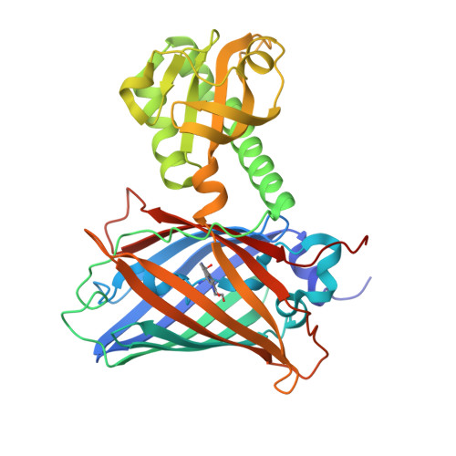
9MRH - PubMed Abstract:
Glycolytic enzymes sense metabolite levels to adapt rapidly to changing energy demands, but measuring the levels of these effectors with spatiotemporal precision in live cells has been challenging. We addressed this question in the context of neuronal depolarization, which activates glycolysis, focusing on the glycolysis inhibitor citrate. We engineered a pair of quantitative fluorescent biosensors for citrate that address several limitations (affinity, pH, Mg 2+ , and temperature) of existing citrate biosensors. Using two-photon fluorescence lifetime imaging, we found that free citrate in the cytosol of neurons in acute mouse brain slices declines two-to-threefold within seconds of neuronal activation and then returns to baseline over a few minutes. The stimulation-dependent citrate transient depends at least in part on the mitochondrial calcium uniporter. These types of live metabolite measurements are essential for achieving a nuanced understanding of the fast control of glycolysis.
- Department of Neurobiology, Harvard Medical School, Boston, MA 02115.
Organizational Affiliation: