9MEX | pdb_00009mex

Structure of phosphocysteine intermediate of human PRL1 phosphatase


Experimental Data Snapshot

  • Method: X-RAY DIFFRACTION
  • Resolution: 2.60 Å
  • R-Value Free: 
    0.237 (Depositor), 0.237 (DCC) 
  • R-Value Work: 
    0.194 (Depositor), 0.194 (DCC) 
  • R-Value Observed: 
    0.196 (Depositor) 

Starting Model: experimental
View more details

wwPDB Validation   3D Report Full Report


This is version 1.2 of the entry. See complete history


Literature

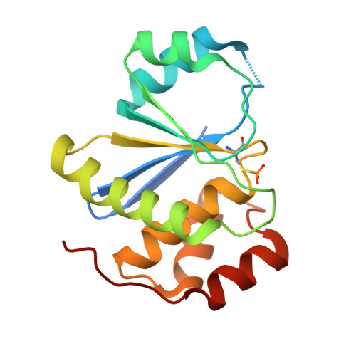
Structure of the phosphocysteine intermediate of the phosphatase of regenerating liver PTP4A1.

Mahbub, L.Kozlov, G.Knorn, C.Gehring, K.

(2025) J Biological Chem 301: 110251-110251

  • DOI: https://doi.org/10.1016/j.jbc.2025.110251
  • Primary Citation of Related Structures:  
    9MEX

  • PubMed Abstract: 

    Phosphatases of regenerating liver (PRL or PTP4A) are protein phosphatases implicated in cell growth, magnesium homeostasis, and cancer metastasis. During catalysis, a phosphocysteine intermediate forms, which must undergo hydrolysis to regenerate the active enzyme. In addition to dephosphorylating substrates, PRLs act as pseudo-phosphatases and bind CBS-pair domain divalent metal cation transport mediators (CNNMs) to regulate magnesium transport. In this study, we investigate the role of PRL residues in phosphocysteine hydrolysis using mutagenesis, enzyme assays, and X-ray crystallography. Loss of an aspartic acid and cysteine in the catalytic site disrupts hydrolysis and stabilizes the phosphocysteine intermediate for weeks. We use this C49S/D72A double mutant to determine the crystal structure of the cysteine phosphorylated form of PRL1 (PTP4A1). The structure confirms that phosphocysteine sterically interferes with CNNM binding, consistent with previous biochemical studies. In vitro enzyme assays reveal the aspartic acid mutation increases the initial rate of catalysis for all three PRL paralogs while the homologous mutation in the phosphatases, PTP1B and PTPN12, disrupts catalysis. This highlights the mechanistic differences between PRLs and classical protein tyrosine phosphatases. Our findings refine our understanding of PRL catalysis and identify novel mutations for investigating PRL function in cancer and magnesium homeostasis.


  • Organizational Affiliation
    • Department of Biochemistry, McGill University, Montreal, QC, H3G0B1, Canada; Centre de Recherche en Biologie Structurale, McGill University, Montreal, QC, H3G0B1, Canada.

Macromolecules
Find similar proteins by:  (by identity cutoff)  |  3D Structure
Entity ID: 1
MoleculeChains Sequence LengthOrganismDetailsImage
Protein tyrosine phosphatase type IVA 1
A, B
164Homo sapiensMutation(s): 2 
Gene Names: PTP4A1PRL1PTPCAAX1
EC: 3.1.3.48
UniProt & NIH Common Fund Data Resources
Find proteins for Q93096 (Homo sapiens)
Explore Q93096 
Go to UniProtKB:  Q93096
PHAROS:  Q93096
GTEx:  ENSG00000112245 
Entity Groups  
Sequence Clusters30% Identity50% Identity70% Identity90% Identity95% Identity100% Identity
UniProt GroupQ93096
Sequence Annotations
Expand
  • Reference Sequence
Experimental Data & Validation

Experimental Data

  • Method: X-RAY DIFFRACTION
  • Resolution: 2.60 Å
  • R-Value Free:  0.237 (Depositor), 0.237 (DCC) 
  • R-Value Work:  0.194 (Depositor), 0.194 (DCC) 
  • R-Value Observed: 0.196 (Depositor) 
Space Group: I 21 3
Unit Cell:
Length ( Å )Angle ( ˚ )
a = 148.164α = 90
b = 148.164β = 90
c = 148.164γ = 90
Software Package:
Software NamePurpose
PHENIXrefinement
HKL-2000data reduction
HKL-2000data scaling
PHENIXphasing

Structure Validation

View Full Validation Report


Entry History & Funding Information

Deposition Data


Funding OrganizationLocationGrant Number
Natural Sciences and Engineering Research Council (NSERC, Canada)CanadaRGPIN-2020-07195

Revision History  (Full details and data files)

  • Version 1.0: 2025-05-14
    Type: Initial release
  • Version 1.1: 2025-06-04
    Changes: Database references
  • Version 1.2: 2025-06-25
    Changes: Database references