Structural and functional constraints on spike activation and host protease utilization limit cell entry of SARS-CoV-2-related bat coronaviruses.
Li, Q., Cai, X., Li, X., Zhang, Y., Li, R., Kang, Z., Wan, D., Wang, J., Li, L., Yang, J., Shi, J., Jin, S., Sun, X., Peng, Y., Zang, N., Xie, Z., Wan, Y., Shang, J.(2025) J Virol 99: e0100725-e0100725
- PubMed: 40704806 Search on PubMed
- DOI: https://doi.org/10.1128/jvi.01007-25
- Primary Citation Related Structures:
9K6Z, 9K75 - PubMed Abstract:
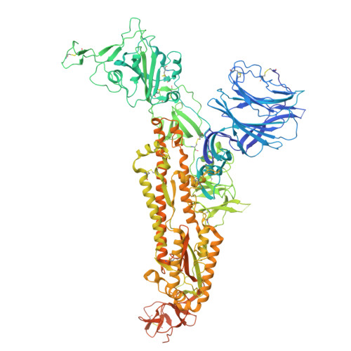
The persistent threat posed by SARS-CoV-2-related coronaviruses (SC2r-CoVs) in wildlife highlights the risk of zoonotic transmission. Cross-species infectivity is predominantly determined by spike (S) character and S-mediated cell entry. In this study, we systematically investigated BANAL-52 and BANAL-103, which exhibit the closest genetic proximity to SARS-CoV-2, focusing on their spike structures and functional characteristics. First, despite comparable receptor-binding domain (RBD)-ACE2 interactions, the spikes of BANAL-52 and BANAL-103 displayed significantly reduced ACE2 binding compared to SARS-CoV-2, suggesting impaired S activation. Second, Cryo-EM structural analyses revealed that BANAL-52 S is stabilized in a "locked" state through linoleic acid (LA) binding and an additional N370 glycan, whereas BANAL-103 S adopts a "closed" conformation due to a unique glycan network. Site-directed mutagenesis targeting the LA binding pocket confirmed that Y365 is related to S conformational transitions and viral entry. Third, both BANAL spikes relied predominantly on lysosomal proteases (e.g., cathepsins) for membrane fusion, unlike SARS-CoV-2, which utilizes a broader range of proteases (e.g., TMPRSS2 and furin). The introduction of a furin cleavage site enhanced the fusogenicity of BANAL spikes. Finally, sera from individuals who have recovered from SARS-CoV-2 effectively neutralized BANAL pseudoviruses, underscoring conserved antigenicity. Our findings elucidate structural and proteolytic barriers that restrict the zoonotic potential of these viruses and propose targeted surveillance strategies to preempt the emergence of SC2r-CoVs. The viral entry mechanisms, which are primarily related to the spike character, play a critical role in determining zoonotic potential. Among the currently identified SC2r-CoVs, BANAL-52 and BANAL-103 exhibit spike proteins with the highest sequence similarity to SARS-CoV-2, rendering them optimal models for comparative studies on S-mediated cell entry and cross-species transmission. In this study, we systematically investigated the molecular constraints governing the functionality of BANAL spikes, with a focus on S-ACE2 interactions, S activation, S structures, and host protease utilization. Notably, we resolved the cryo-EM structure of BANAL-52 S at neutral pH and the first cryo-EM structure of BANAL-103 S, revealing distinct glycan- and lipid-mediated stabilization of inactive states. Furthermore, cross-neutralization assays demonstrated that sera of convalescents from SARS-CoV-2 inhibited BANAL pseudovirus entry with an efficiency of approximately 80%, thereby highlighting conserved antigenic epitopes and informing the development of broad-spectrum therapeutic strategies against emerging SC2r-CoVs.
- Henan Institute of Medical and Pharmaceutical Sciences, Zhengzhou University, Zhengzhou, Henan, China.
Organizational Affiliation: