9JYW | pdb_00009jyw

Crystal structure of the gamma-carbonic anhydrase from the polyextremophilic bacterium Aeribacillus pallidus


Experimental Data Snapshot

  • Method: X-RAY DIFFRACTION
  • Resolution: 1.70 Å
  • R-Value Free: 
    0.245 (Depositor), 0.225 (DCC) 
  • R-Value Work: 
    0.213 (Depositor), 0.205 (DCC) 
  • R-Value Observed: 
    0.215 (Depositor) 

Starting Model: experimental
View more details

wwPDB Validation 3D Report Full Report

Validation slider image for 9JYW

Ligand Structure Quality Assessment 


This is version 1.2 of the entry. See complete history

Literature

Crystal structure of gamma-carbonic anhydrase from the polyextremophilic bacterium Aeribacillus pallidus.

Choi, S.H.Jin, M.S.

(2024) Mol Cells 48: 100165-100165

  • DOI: https://doi.org/10.1016/j.mocell.2024.100165
  • Primary Citation Related Structures: 
    9JYW

  • PubMed Abstract: 

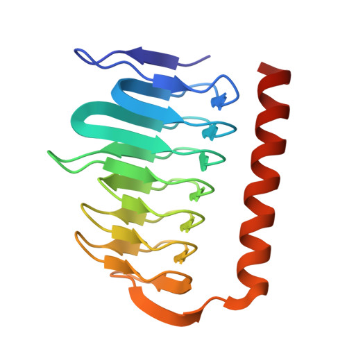
    The polyextremophilic bacterium Aeribacillus pallidus produces a thermo- and alkali-stable γ-carbonic anhydrase (γ-apCA), a homotrimeric metalloenzyme containing a zinc ion in its active site that catalyzes the reversible hydration of carbon dioxide (CO 2 ). Here, we present the first crystal structure of γ-apCA at 1.7 Å resolution, revealing two trimers in the asymmetric unit. The overall structure is consistent with other γ-CAs, where each monomer adopts a prism-like structure consisting of an N-terminal left-handed β-helix and a C-terminal α-helix. The active site, located at the interface between two monomers, coordinates the zinc ion with three histidine residues (H65, H82, and H87) and a water molecule in a tetrahedral configuration. Structural comparison indicates that the amino acid composition at the active site of γ-apCA differs significantly from the prototypic γ-CA from Methanosarcina thermophila. This variation likely accounts for the lack of measurable CO 2 hydration activity in γ-apCA. Additionally, the structure reveals non-catalytic zinc and sulfate ions trapped at the trimer core and trimer-trimer non-crystallographic interfaces. These may contribute to stabilizing enzyme assembly and promoting crystal packing.


  • Organizational Affiliation
    • School of Life Sciences, GIST, 123 Cheomdangwagi-ro, Buk-gu, Gwangju 61005, Republic of Korea.

Macromolecule Content 

  • Total Structure Weight: 117.14 kDa 
  • Atom Count: 8,550 
  • Modeled Residue Count: 1,026 
  • Deposited Residue Count: 1,050 
  • Unique protein chains: 1

Macromolecules

Find similar proteins by:|  3D Structure
Entity ID: 1
MoleculeChains  Sequence LengthOrganismDetailsImage
Gamma carbonic anhydrase family protein
A, B, C, D, E
A, B, C, D, E, F
175Aeribacillus pallidusMutation(s): 0 
Gene Names: AP3564_09575
UniProt
Find proteins for A0A223E5F5 (Aeribacillus pallidus)
Explore A0A223E5F5 
Go to UniProtKB:  A0A223E5F5
Entity Groups
Sequence Clusters30% Identity50% Identity70% Identity90% Identity95% Identity100% Identity
UniProt GroupA0A223E5F5
Sequence Annotations
Expand
Reference Sequence

Small Molecules

Ligands 2 Unique
IDChains Name / Formula / InChI Key2D Diagram3D Interactions
SO4
(Subject of Investigation/LOI)

Query on SO4


Download:Ideal Coordinates CCD File
M [auth B],
R [auth D]
SULFATE ION
O4 S
QAOWNCQODCNURD-UHFFFAOYSA-L
ZN
(Subject of Investigation/LOI)

Query on ZN


Download:Ideal Coordinates CCD File
G [auth A]
H [auth A]
I [auth B]
J [auth B]
K [auth B]
G [auth A],
H [auth A],
I [auth B],
J [auth B],
K [auth B],
L [auth B],
N [auth C],
O [auth D],
P [auth D],
Q [auth D],
S [auth E],
T [auth F]
ZINC ION
Zn
PTFCDOFLOPIGGS-UHFFFAOYSA-N

Experimental Data & Validation

Experimental Data

  • Method: X-RAY DIFFRACTION
  • Resolution: 1.70 Å
  • R-Value Free:  0.245 (Depositor), 0.225 (DCC) 
  • R-Value Work:  0.213 (Depositor), 0.205 (DCC) 
  • R-Value Observed: 0.215 (Depositor) 
Space Group: C 1 2 1
Unit Cell:
Length ( Å )Angle ( ˚ )
a = 118.911α = 90
b = 110.672β = 118.8
c = 98.578γ = 90
Software Package:
Software NamePurpose
REFMACrefinement
HKL-2000data scaling
HKL-2000data reduction
PHASERphasing

Structure Validation

View Full Validation Report


Ligand Structure Quality Assessment 


Entry History 

& Funding Information

Deposition Data


Funding OrganizationLocationGrant Number
National Research Foundation (NRF, Korea)Korea, Republic OfNRF-2022R1A2C1091278
National Research Foundation (NRF, Korea)Korea, Republic OfRS-2024-00344154

Revision History  (Full details and data files)

  • Version 1.0: 2024-12-11
    Type: Initial release
  • Version 1.1: 2024-12-18
    Changes: Database references
  • Version 1.2: 2025-01-01
    Changes: Database references