9JUB | pdb_00009jub

Cryo-EM structure of human hyaluronidase PH-20


Experimental Data Snapshot

  • Method: ELECTRON MICROSCOPY
  • Resolution: 3.10 Å
  • Aggregation State: PARTICLE 
  • Reconstruction Method: SINGLE PARTICLE 

wwPDB Validation 3D Report Full Report

Validation slider image for 9JUB

This is version 1.3 of the entry. See complete history

Literature

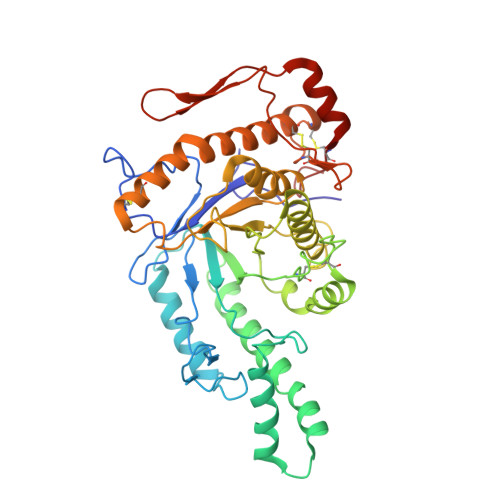
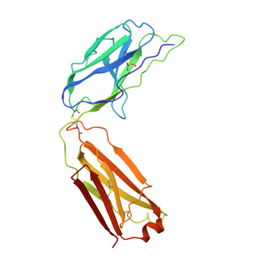
Cryo-EM Structure of Human Hyaluronidase PH-20.

Im, S.B.Song, H.N.Jeong, T.K.Kim, N.Kim, K.Park, S.J.Oh, B.H.

(2025) Proteins 93: 1067-1073

  • DOI: https://doi.org/10.1002/prot.26788
  • Primary Citation Related Structures: 
    9JUB

  • PubMed Abstract: 

    PH-20 is a specific type of hyaluronidase that plays a critical role in the fertilization process by facilitating the initial binding of sperm to the glycoprotein layer surrounding the oocyte and subsequently breaking down hyaluronic acid polymers in the cumulus cell layer. PH-20 contains an epidermal growth factor (EGF)-like domain, which may be involved in the recognition of the glycoprotein layer in addition to the catalytic domain. Herein, we report the structure of human PH-20 determined by cryogenic electron microscopy. Comparative analyses of the PH-20 structure with two other available hyaluronidase structures reveal a general similarity in the central catalytic domains, including the conservation of catalytically essential residues at the equivalent spatial positions. However, unique difference is found in the EGF-like domain, characterized by a longer sequence that is likely to form a flexibly anchored β-hairpin containing a disulfide bond.


  • Organizational Affiliation
    • Department of Biological Sciences, KAIST Institute for the Biocentury, Korea Advanced Institute of Science and Technology, Daejeon, Republic of Korea.

Macromolecule Content 

  • Total Structure Weight: 92.63 kDa 
  • Atom Count: 6,479 
  • Modeled Residue Count: 829 
  • Deposited Residue Count: 834 
  • Unique protein chains: 3

Macromolecules

Find similar proteins by:|  3D Structure
Entity ID: 1
MoleculeChains  Sequence LengthOrganismDetailsImage
Hyaluronidase PH-20406Homo sapiensMutation(s): 0 
Gene Names: SPAM1HYAL3PH20
EC: 3.2.1.35
UniProt & NIH Common Fund Data Resources
Find proteins for P38567 (Homo sapiens)
Explore P38567 
Go to UniProtKB:  P38567
GTEx:  ENSG00000106304 
Entity Groups
Sequence Clusters30% Identity50% Identity70% Identity90% Identity95% Identity100% Identity
UniProt GroupP38567
Glycosylation
Glycosylation Sites: 3Go to GlyGen: P38567-1
Sequence Annotations
Expand
Reference Sequence
Find similar proteins by:|  3D Structure
Entity ID: 2
MoleculeChains  Sequence LengthOrganismDetailsImage
Heavy chain of 79C11Fab216Oryctolagus cuniculusMutation(s): 0 
Find similar proteins by:|  3D Structure
Entity ID: 3
MoleculeChains  Sequence LengthOrganismDetailsImage
Light chain of 79C11Fab212Oryctolagus cuniculusMutation(s): 0 

Oligosaccharides

Help  
Entity ID: 4
MoleculeChains Length2D Diagram GlycosylationD Interactions
2-acetamido-2-deoxy-beta-D-glucopyranose-(1-4)-2-acetamido-2-deoxy-beta-D-glucopyranose
D
2N-Glycosylation
Glycosylation Resources
GlyTouCan: G42666HT
GlyCosmos: G42666HT
GlyGen: G42666HT

Experimental Data & Validation

Experimental Data

  • Method: ELECTRON MICROSCOPY
  • Resolution: 3.10 Å
  • Aggregation State: PARTICLE 
  • Reconstruction Method: SINGLE PARTICLE 

Structure Validation

View Full Validation Report


Entry History 

& Funding Information

Deposition Data


Funding OrganizationLocationGrant Number
Other governmentKorea, Republic Of2023R1A2C3007329
Other governmentKorea, Republic OfNRF-2021M3A9G8025599

Revision History  (Full details and data files)

  • Version 1.0: 2024-10-30
    Type: Initial release
  • Version 1.1: 2024-11-20
    Changes: Data collection
  • Version 1.2: 2025-01-15
    Changes: Data collection, Database references
  • Version 1.3: 2025-04-16
    Changes: Data collection, Database references