9JTE | pdb_00009jte

Crystal structure of SARS-CoV-2 receptor binding domain complexed with fox ACE2


Experimental Data Snapshot

  • Method: X-RAY DIFFRACTION
  • Resolution: 3.66 Å
  • R-Value Free: 
    0.249 (Depositor), 0.249 (DCC) 
  • R-Value Work: 
    0.228 (Depositor), 0.231 (DCC) 
  • R-Value Observed: 
    0.229 (Depositor) 

Starting Model: experimental
View more details

wwPDB Validation 3D Report Full Report

Validation slider image for 9JTE

Ligand Structure Quality Assessment 


This is version 1.1 of the entry. See complete history

Literature

Molecular basis of fox ACE2 recognition by receptor binding domains of SARS-CoV-2 and PCoV-GD.

Wang, C.Nan, X.Pei, C.Li, M.Wu, J.Wang, Z.Fan, S.Lan, J.

(2026) Cell Insight 5: 100314-100314

  • DOI: https://doi.org/10.1016/j.cellin.2026.100314
  • Primary Citation Related Structures: 
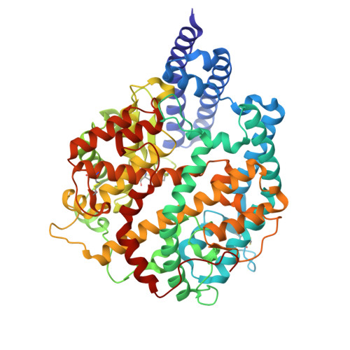
    9JTD, 9JTE

  • PubMed Abstract: 

    The severe acute respiratory syndrome coronavirus 2 (SARS-CoV-2) positive RNA detected in red fox swab samples provide evidence of a natural SARS-CoV-2 infection in this species. Many studies have also demonstrated that the red fox angiotensin converting enzyme 2 (fACE2) could serve as the receptor of SARS-CoV-2 and many SARS-CoV-2 related sarbecoviruses. However, the molecular mechanisms of fACE2 attached by animal-origin coronaviruses remain poorly understood. Here, we found that fACE2 could mediate pseudovirus entry and cell membrane fusion induced by the SARS-CoV-2 and pangolin coronavirus isolated from Guangdong (PCoV-GD) spikes as human ACE2 (hACE2). The binding affinity of fACE2 bound to the receptor-binding domain (RBD) of PCoV-GD was stronger than the SARS-CoV-2 RBD demonstrated by surface plasmon resonance (SPR) assay, while deglycosylation of PCoV-GD RBD N370 by T372A mutation or glycosylation of SARS-CoV-2 RBD N370 by A372T mutation had little effect on fACE2 binding compared with wild-type (WT) RBD. We further solved the crystal structures of fACE2 bound to SARS-CoV-2 RBD and PCoV-GD T372A mutant RBD. Interface analysis and SPR assay revealed that the R417 and H498 of PCoV-GD RBD might account for the binding affinity enhancement with fACE2 compared with K417 and Q498 of SARS-CoV-2 RBD. Moreover, the Y453F mutation in SARS-CoV-2 RBD increased the binding affinity to fACE2 while this mutation in PCoV-GD RBD decreased the binding affinity to fACE2 compared with the corresponding WT RBDs. Furthermore, the RBDs of many prevalent SARS-CoV-2 variants could all bind to fACE2. Our results indicated that the continuous surveillance of SARS-CoV-2 and related sarbecoviruses in fox species was also necessary to better prevent animal-to-human spillover of the coronaviruses.


  • Organizational Affiliation
    • School of Biomedical Sciences, Hunan University, Changsha, Hunan, China.

Macromolecule Content 

  • Total Structure Weight: 184.36 kDa 
  • Atom Count: 12,947 
  • Modeled Residue Count: 1,571 
  • Deposited Residue Count: 1,574 
  • Unique protein chains: 2

Macromolecules

Find similar proteins by:|  3D Structure
Entity ID: 1
MoleculeChains  Sequence LengthOrganismDetailsImage
Angiotensin-converting enzyme
A, B
592Vulpes vulpesMutation(s): 0 
Gene Names: ACE2
EC: 3.4
UniProt
Find proteins for A0A3Q7RAT9 (Vulpes vulpes)
Explore A0A3Q7RAT9 
Go to UniProtKB:  A0A3Q7RAT9
Entity Groups
Sequence Clusters30% Identity50% Identity70% Identity90% Identity95% Identity100% Identity
UniProt GroupA0A3Q7RAT9
Glycosylation
Glycosylation Sites: 3
Sequence Annotations
Expand
Reference Sequence
Find similar proteins by:|  3D Structure
Entity ID: 2
MoleculeChains  Sequence LengthOrganismDetailsImage
Spike protein S1C [auth E],
D [auth C]
195Severe acute respiratory syndrome coronavirus 2Mutation(s): 0 
Gene Names: S2
UniProt
Find proteins for P0DTC2 (Severe acute respiratory syndrome coronavirus 2)
Explore P0DTC2 
Go to UniProtKB:  P0DTC2
Entity Groups
Sequence Clusters30% Identity50% Identity70% Identity90% Identity95% Identity100% Identity
UniProt GroupP0DTC2
Glycosylation
Glycosylation Sites: 1Go to GlyGen: P0DTC2-1
Sequence Annotations
Expand
Reference Sequence

Oligosaccharides

Help  
Entity ID: 3
MoleculeChains Length2D Diagram GlycosylationD Interactions
beta-D-mannopyranose-(1-4)-2-acetamido-2-deoxy-beta-D-glucopyranose-(1-4)-2-acetamido-2-deoxy-beta-D-glucopyranoseE [auth D]3N-Glycosylation
Glycosylation Resources
GlyTouCan: G15407YE
GlyCosmos: G15407YE
GlyGen: G15407YE
Entity ID: 4
MoleculeChains Length2D Diagram GlycosylationD Interactions
2-acetamido-2-deoxy-beta-D-glucopyranose-(1-4)-2-acetamido-2-deoxy-beta-D-glucopyranose
F, G, H
2N-Glycosylation
Glycosylation Resources
GlyTouCan: G42666HT
GlyCosmos: G42666HT
GlyGen: G42666HT

Experimental Data & Validation

Experimental Data

  • Method: X-RAY DIFFRACTION
  • Resolution: 3.66 Å
  • R-Value Free:  0.249 (Depositor), 0.249 (DCC) 
  • R-Value Work:  0.228 (Depositor), 0.231 (DCC) 
  • R-Value Observed: 0.229 (Depositor) 
Space Group: I 21 21 21
Unit Cell:
Length ( Å )Angle ( ˚ )
a = 149.976α = 90
b = 192.714β = 90
c = 194.791γ = 90
Software Package:
Software NamePurpose
PHENIXrefinement
HKL-3000data reduction
HKL-3000data scaling
PHENIXphasing

Structure Validation

View Full Validation Report


Ligand Structure Quality Assessment 


Entry History 

& Funding Information

Deposition Data


Funding OrganizationLocationGrant Number
Not funded--

Revision History  (Full details and data files)

  • Version 1.0: 2025-10-22
    Type: Initial release
  • Version 1.1: 2026-05-06
    Changes: Database references