9ISK | pdb_00009isk

Cryo-EM structure of KpFtsZ-ZapA complex


Experimental Data Snapshot

  • Method: ELECTRON MICROSCOPY
  • Resolution: 2.73 Å
  • Aggregation State: FILAMENT 
  • Reconstruction Method: HELICAL 

Starting Models: experimental
View more details

wwPDB Validation 3D Report Full Report

Validation slider image for 9ISK

This is version 1.0 of the entry. See complete history

Literature

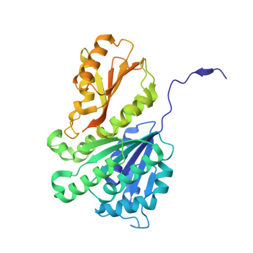
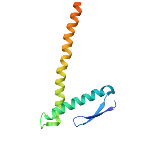
Structural basis for the interaction between the bacterial cell division proteins FtsZ and ZapA.

Fujita, J.Kasai, K.Hibino, K.Kagoshima, G.Kamimura, N.Tobita, S.Kato, Y.Uehara, R.Namba, K.Uchihashi, T.Matsumura, H.

(2025) Nat Commun 16: 5985-5985

  • DOI: https://doi.org/10.1038/s41467-025-60940-w
  • Primary Citation Related Structures: 
    9ISJ, 9ISK

  • PubMed Abstract: 

    Cell division in most bacteria is regulated by the tubulin homolog FtsZ as well as ZapA, a FtsZ-associated protein. However, how FtsZ and ZapA function coordinately has remained elusive. Here we report the cryo-electron microscopy structure of the ZapA-FtsZ complex at 2.73 Å resolution. The complex forms an asymmetric ladder-like structure, in which the double antiparallel FtsZ protofilament on one side and a single protofilament on the other side are tethered by ZapA tetramers. In the complex, the extensive interactions of FtsZ with ZapA cause a structural change of the FtsZ protofilament, and the formation of the double FtsZ protofilament increases electrostatic repulsion. High-speed atomic force microscopy analysis revealed cooperative interactions of ZapA with FtsZ at a molecular level. Our findings not only provide a structural basis for the interaction between FtsZ and ZapA but also shed light on how ZapA binds to FtsZ protofilaments without disturbing FtsZ dynamics to promote cell division.


  • Organizational Affiliation
    • Graduate School of Frontier Biosciences, University of Osaka, Osaka, Japan.

Macromolecule Content 

  • Total Structure Weight: 346.66 kDa 
  • Atom Count: 17,394 
  • Modeled Residue Count: 2,324 
  • Deposited Residue Count: 3,170 
  • Unique protein chains: 2

Macromolecules

Find similar proteins by:|  3D Structure
Entity ID: 1
MoleculeChains  Sequence LengthOrganismDetailsImage
Cell division protein FtsZ
A, B, C, D, E
A, B, C, D, E, F
383Klebsiella pneumoniae subsp. pneumoniae MGH 78578Mutation(s): 0 
Gene Names: ftsZKPN_00099
UniProt
Find proteins for A6T4N8 (Klebsiella pneumoniae subsp. pneumoniae (strain ATCC 700721 / MGH 78578))
Explore A6T4N8 
Go to UniProtKB:  A6T4N8
Entity Groups
Sequence Clusters30% Identity50% Identity70% Identity90% Identity95% Identity100% Identity
UniProt GroupA6T4N8
Sequence Annotations
Expand
Reference Sequence
Find similar proteins by:|  3D Structure
Entity ID: 2
MoleculeChains  Sequence LengthOrganismDetailsImage
Cell division protein ZapA109Klebsiella variicola 342Mutation(s): 0 
Gene Names: zapAKPK_0754
UniProt
Find proteins for B5XUC8 (Klebsiella variicola (strain 342))
Explore B5XUC8 
Go to UniProtKB:  B5XUC8
Entity Groups
Sequence Clusters30% Identity50% Identity70% Identity90% Identity95% Identity100% Identity
UniProt GroupB5XUC8
Sequence Annotations
Expand
Reference Sequence

Small Molecules

Ligands 3 Unique
IDChains Name / Formula / InChI Key2D Diagram3D Interactions
G2P
(Subject of Investigation/LOI)

Query on G2P


Download:Ideal Coordinates CCD File
AA [auth E]
DA [auth F]
O [auth A]
R [auth B]
U [auth C]
AA [auth E],
DA [auth F],
O [auth A],
R [auth B],
U [auth C],
X [auth D]
PHOSPHOMETHYLPHOSPHONIC ACID GUANYLATE ESTER
C11 H18 N5 O13 P3
GXTIEXDFEKYVGY-KQYNXXCUSA-N
K
(Subject of Investigation/LOI)

Query on K


Download:Ideal Coordinates CCD File
CA [auth E]
FA [auth F]
Q [auth A]
T [auth B]
W [auth C]
CA [auth E],
FA [auth F],
Q [auth A],
T [auth B],
W [auth C],
Z [auth D]
POTASSIUM ION
K
NPYPAHLBTDXSSS-UHFFFAOYSA-N
MG
(Subject of Investigation/LOI)

Query on MG


Download:Ideal Coordinates CCD File
BA [auth E]
EA [auth F]
P [auth A]
S [auth B]
V [auth C]
BA [auth E],
EA [auth F],
P [auth A],
S [auth B],
V [auth C],
Y [auth D]
MAGNESIUM ION
Mg
JLVVSXFLKOJNIY-UHFFFAOYSA-N

Experimental Data & Validation

Experimental Data

  • Method: ELECTRON MICROSCOPY
  • Resolution: 2.73 Å
  • Aggregation State: FILAMENT 
  • Reconstruction Method: HELICAL 
EM Software:
TaskSoftware PackageVersion
RECONSTRUCTIONcryoSPARC4.2.1
MODEL REFINEMENTPHENIX1.19.2

Structure Validation

View Full Validation Report


Entry History 

& Funding Information

Deposition Data


Funding OrganizationLocationGrant Number
Japan Society for the Promotion of Science (JSPS)JapanJP20K22630
Japan Society for the Promotion of Science (JSPS)JapanJP23K06418
Japan Society for the Promotion of Science (JSPS)JapanJP24K01994
Japan Society for the Promotion of Science (JSPS)JapanJP24H02277
Japan Society for the Promotion of Science (JSPS)JapanJP24H02270
Japan Society for the Promotion of Science (JSPS)JapanJP23K18033
Japan Science and TechnologyJapanJPMJOP1861
Japan Agency for Medical Research and Development (AMED)JapanJP21am0101117
Japan Agency for Medical Research and Development (AMED)JapanJP22ama121003
Japan Agency for Medical Research and Development (AMED)JapanJP23ama121001
Japan Agency for Medical Research and Development (AMED)JapanJP17pc0101020

Revision History  (Full details and data files)

  • Version 1.0: 2025-07-23
    Type: Initial release