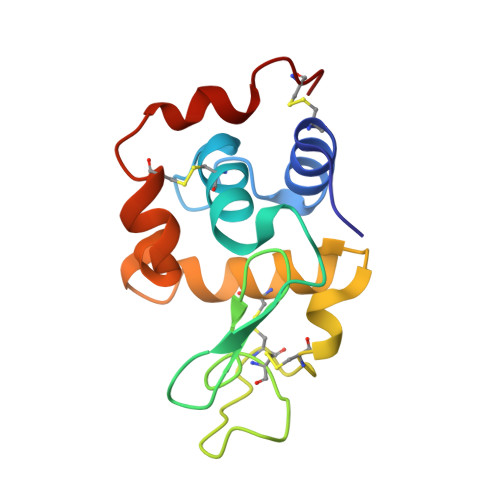
9IG6 | pdb_00009ig6

Hen egg-white lysozyme structure determined by 3DED/MicroED on a 200 keV microscope


Experimental Data Snapshot

  • Method: ELECTRON CRYSTALLOGRAPHY
  • Resolution: 2.80 Å
  • R-Value Free: 
    0.326 (Depositor), 0.360 (DCC) 
  • R-Value Work: 
    0.294 (Depositor), 0.306 (DCC) 

Starting Model: experimental
View more details

wwPDB Validation 3D Report Full Report

Validation slider image for 9IG6

This is version 1.1 of the entry. See complete history. 

Literature

Strategies for mitigating radiation damage and improving data completeness in 3D electron diffraction of protein crystals.

Shaikhqasem, A., Hamdi, F., Machner, L., Parthier, C., Breithaupt, C., Kyrilis, F.L., Feller, S.M., Kastritis, P.L., Stubbs, M.T.

(2026) Acta Crystallogr D Struct Biol 82: 11-22

  • DOI: https://doi.org/10.1107/S2059798325011258
  • Primary Citation Related Structures: 
    9IG6

  • PubMed Abstract: 

    While 3D electron diffraction (3D-ED or microcrystal electron diffraction; MicroED) has emerged as a promising method for protein structure determination, its applicability is hindered by a high susceptibility to radiation damage, leading to a decreasing signal-to-noise ratio in consecutive diffraction patterns that limits the quality (resolution and redundancy) of the data. In addition, data completeness may be restricted due to the geometrical limitations of current sample holders and stages. Although specialized equipment can overcome these challenges, many laboratories do not have access to such instrumentation. In this work, we introduce an approach that addresses these issues using a commonly available 200 keV cryo-electron microscope. The multi-position acquisition technique that we present here combines (i) multiple data acquisitions from a single crystal over several tilt ranges and (ii) merging data from a small number of crystals each tilted about a different axis. The robustness of this approach is demonstrated by the de novo elucidation of a protein-peptide complex structure from only two orthorhombic microcrystals.


  • Organizational Affiliation: 
    • Charles-Tanford-Proteinzentrum, Martin-Luther-Universität Halle-Wittenberg, Halle (Saale), Germany.

Macromolecule Content 

  • Total Structure Weight: 14.33 kDa 
  • Atom Count: 1,013 
  • Modeled Residue Count: 129 
  • Deposited Residue Count: 129 
  • Unique protein chains: 1

Macromolecules

Find similar proteins by:|  3D Structure
Entity ID: 1
MoleculeChains  Sequence LengthOrganismDetailsImage
Lysozyme C129Gallus gallusMutation(s): 0 
EC: 3.2.1.17
UniProt
Find proteins for P00698 (Gallus gallus)
Explore P00698 
Go to UniProtKB:  P00698
Entity Groups
Sequence Clusters30% Identity50% Identity70% Identity90% Identity95% Identity100% Identity
UniProt GroupP00698
Sequence Annotations
Expand
Reference Sequence

Experimental Data & Validation

Experimental Data

  • Method: ELECTRON CRYSTALLOGRAPHY
  • Resolution: 2.80 Å
  • R-Value Free:  0.326 (Depositor), 0.360 (DCC) 
  • R-Value Work:  0.294 (Depositor), 0.306 (DCC) 
Space Group: P 43 21 2
Unit Cell:
Length ( Å )Angle ( ˚ )
a = 78.59α = 90
b = 78.59β = 90
c = 37.84γ = 90
Software Package:
Software NamePurpose
XDSdata scaling
REFMACrefinement

Structure Validation

View Full Validation Report


Entry History 

& Funding Information

Deposition Data


Funding OrganizationLocationGrant Number
Other governmentBMBF ZIK HALOmem, grant no. 03Z22HN23
Other governmentBMBF ZIK HALOmem, grant no. 03Z22HI2
Other governmentBMBF, CORONAmem, grant no. 03COV04
Other governmentEFRE for Saxony-Anhalt (grant no. ZS/2016/04/78115 and grant no. ZS/2024/05/187255
Other governmentDFG, project numbers 391498659, RTG 2467 and 514901783, CRC 1664
Other governmentThe European Union Horizon Europe ERA Chair hot4cryo, project number 101086665

Revision History  (Full details and data files)

  • Version 1.0: 2025-03-19
    Type: Initial release
  • Version 1.1: 2026-09-30
    Changes: Database references