9I58 | pdb_00009i58

Crystal structure of DNPH1 bound by compound 31.


Experimental Data Snapshot

  • Method: X-RAY DIFFRACTION
  • Resolution: 1.21 Å
  • R-Value Free: 
    0.233 (Depositor), 0.225 (DCC) 
  • R-Value Work: 
    0.203 (Depositor), 0.192 (DCC) 
  • R-Value Observed: 
    0.204 (Depositor) 

Starting Model: experimental
View more details

wwPDB Validation   3D Report Full Report


Ligand Structure Quality Assessment 


This is version 1.1 of the entry. See complete history


Literature

Discovery and Optimization of a Non-Nucleoside-Based Series of Inhibitors of 2'-Deoxynucleoside 5'-Monophosphate Glycosidase (DNPH1).

Barlaam, B.Alonso-Crisostomo, L.Anderson, N.A.Argyrou, A.Astles, P.C.Cadogan, E.B.Carlino, L.Collie, G.W.Davies, N.L.Hall, J.Kitching, L.Li, X.Michopoulos, F.Milbradt, A.G.Nikkila, J.Northall, S.O'Connor, M.J.Pei, X.Shaw, J.Slade, D.Southgate, H.Stead, D.Stubbs, C.J.Whitehurst, B.C.Xing, B.Yuan, Y.Zhou, J.

(2025) J Med Chem 68: 24381-24403

  • DOI: https://doi.org/10.1021/acs.jmedchem.5c02356
  • Primary Citation of Related Structures:  
    9I3Q, 9I57, 9I58, 9I6E

  • PubMed Abstract: 

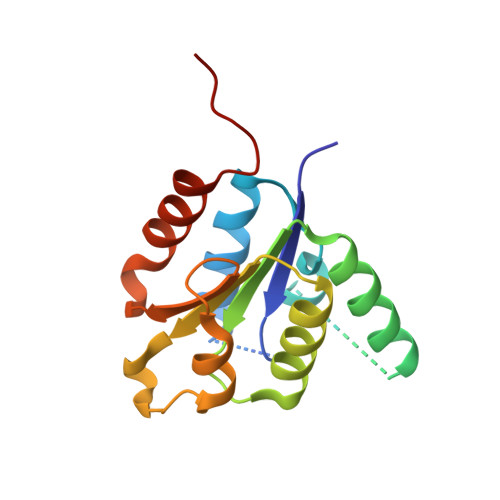
    DNPH1 is a nucleotide pool sanitizer that cleaves 5-hydroxymethyl-2-deoxyuridine-5-monophosphate (hmdUMP), preventing incorporation of the correspondent non-natural nucleotide into DNA. Recent findings have demonstrated that loss of DNPH1 could potentiate the sensitivity of PARP inhibitors in homologous recombination repair (HRR)-deficient cancers. We report the optimization of a non-nucleoside-based series of DNPH1 inhibitors. Starting from a weak compound 1 (binding affinity pIC 50 4.7), we identified compound 38 as a very potent inhibitor of DNPH1 (pIC 50 9.3) using DNPH1 X-ray structure-guided drug design. Compound 38 demonstrated target engagement of DNPH1 in the SUM149PT cell line (pIC 50 7.2). Using this tool compound, we then report the in vitro pharmacology of a DNPH1 inhibitor in the BRCA1 mutant SUM149PT cell line.


  • Organizational Affiliation
    • Oncology, R&D, AstraZeneca, Cambridge CB2 0AA, U.K.

Macromolecules
Find similar proteins by:  (by identity cutoff)  |  3D Structure
Entity ID: 1
MoleculeChains Sequence LengthOrganismDetailsImage
5-hydroxymethyl-dUMP N-hydrolase
A, B
152Homo sapiensMutation(s): 0 
Gene Names: DNPH1C6orf108RCL
EC: 3.2.2
UniProt & NIH Common Fund Data Resources
Find proteins for O43598 (Homo sapiens)
Explore O43598 
Go to UniProtKB:  O43598
PHAROS:  O43598
GTEx:  ENSG00000112667 
Entity Groups  
Sequence Clusters30% Identity50% Identity70% Identity90% Identity95% Identity100% Identity
UniProt GroupO43598
Sequence Annotations
Expand
  • Reference Sequence
Small Molecules
Ligands 1 Unique
IDChains Name / Formula / InChI Key2D Diagram3D Interactions
A1IZX (Subject of Investigation/LOI)
Query on A1IZX

Download Ideal Coordinates CCD File 
C [auth A],
D [auth B]
3-[2-fluoranyl-3-(trifluoromethyl)phenyl]-4-[[4-(methylamino)-6-propan-2-yl-quinazolin-2-yl]amino]benzoic acid
C26 H22 F4 N4 O2
NVXLCRYRBAZDFU-UHFFFAOYSA-N
Experimental Data & Validation

Experimental Data

  • Method: X-RAY DIFFRACTION
  • Resolution: 1.21 Å
  • R-Value Free:  0.233 (Depositor), 0.225 (DCC) 
  • R-Value Work:  0.203 (Depositor), 0.192 (DCC) 
  • R-Value Observed: 0.204 (Depositor) 
Space Group: P 1
Unit Cell:
Length ( Å )Angle ( ˚ )
a = 36.686α = 92.53
b = 39.763β = 99.55
c = 48.28γ = 113.45
Software Package:
Software NamePurpose
BUSTERrefinement
autoPROCdata reduction
autoPROCdata scaling
PHASERphasing

Structure Validation

View Full Validation Report


Ligand Structure Quality Assessment 


Entry History & Funding Information

Deposition Data

  • Released Date: 2025-11-12 
  • Deposition Author(s): Collie, G.W.

Funding OrganizationLocationGrant Number
Other private--

Revision History  (Full details and data files)

  • Version 1.0: 2025-11-12
    Type: Initial release
  • Version 1.1: 2025-12-10
    Changes: Database references