9ELI | pdb_00009eli

Cryo-EM structure of SARS-CoV-2 Omicron KP.3.1.1 spike protein (closed state)


Experimental Data Snapshot

  • Method: ELECTRON MICROSCOPY
  • Resolution: 2.82 Å
  • Aggregation State: PARTICLE 
  • Reconstruction Method: SINGLE PARTICLE 

wwPDB Validation 3D Report Full Report

Validation slider image for 9ELI

This is version 1.1 of the entry. See complete history

Literature

Structural and functional insights into the evolution of SARS-CoV-2 KP.3.1.1 spike protein.

Feng, Z.Huang, J.Baboo, S.Diedrich, J.K.Bangaru, S.Paulson, J.C.Yates 3rd, J.R.Yuan, M.Wilson, I.A.Ward, A.B.

(2025) Cell Rep 44: 115941-115941

  • DOI: https://doi.org/10.1016/j.celrep.2025.115941
  • Primary Citation Related Structures: 
    9ELE, 9ELF, 9ELG, 9ELH, 9ELI, 9ELJ, 9ELK, 9ELL, 9ELM, 9ELN, 9ELO, 9ELP, 9ELQ

  • PubMed Abstract: 

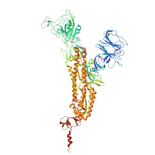
    The JN.1-sublineage KP.3.1.1 recently emerged as the globally prevalent SARS-CoV-2 variant, demonstrating increased infectivity and antibody escape. We investigate how mutations and a deletion in the KP.3.1.1 spike protein (S) affect hACE2 binding and antibody escape. Mass spectrometry confirms a new glycan site at residue N30 that alters the glycoforms at neighboring N61. Cryoelectron microscopy (cryo-EM) structures show that the N30 glycan and rearrangement of adjacent residues do not significantly change the overall spike structure, up-down ratio of receptor-binding domains (RBDs), or hACE2 binding. Furthermore, a KP.3.1.1 S with hACE2 structure further confirms an epistatic effect between F456L and Q493E on hACE2 binding. Our analysis shows that SARS-CoV-2 variants that emerged after late 2023 are now incorporating reversions to residues found in other sarbecoviruses, including the N30 glycan, Q493E, and others. Overall, these results inform on the structural and functional consequences of the KP.3.1.1 mutations, the current SARS-CoV-2 evolutionary trajectory, and immune evasion.


  • Organizational Affiliation
    • Department of Integrative Structural and Computational Biology, The Scripps Research Institute, La Jolla, CA, USA.

Macromolecule Content 

  • Total Structure Weight: 411.52 kDa 
  • Atom Count: 25,509 
  • Modeled Residue Count: 3,153 
  • Deposited Residue Count: 3,597 
  • Unique protein chains: 1

Macromolecules

Find similar proteins by:|  3D Structure
Entity ID: 1
MoleculeChains  Sequence LengthOrganismDetailsImage
Spike glycoprotein
A, B, C
1,199Severe acute respiratory syndrome coronavirus 2Mutation(s): 30 
Gene Names: S2
UniProt
Find proteins for P0DTC2 (Severe acute respiratory syndrome coronavirus 2)
Explore P0DTC2 
Go to UniProtKB:  P0DTC2
Entity Groups
Sequence Clusters30% Identity50% Identity70% Identity90% Identity95% Identity100% Identity
UniProt GroupP0DTC2
Glycosylation
Glycosylation Sites: 12Go to GlyGen: P0DTC2-1
Sequence Annotations
Expand
Reference Sequence

Oligosaccharides

Help  
Entity ID: 2
MoleculeChains Length2D Diagram GlycosylationD Interactions
beta-D-mannopyranose-(1-4)-2-acetamido-2-deoxy-beta-D-glucopyranose-(1-4)-2-acetamido-2-deoxy-beta-D-glucopyranose
D, H, I, M, N
D, H, I, M, N, R
3N-Glycosylation
Glycosylation Resources
GlyTouCan: G15407YE
GlyCosmos: G15407YE
GlyGen: G15407YE
Entity ID: 3
MoleculeChains Length2D Diagram GlycosylationD Interactions
2-acetamido-2-deoxy-beta-D-glucopyranose-(1-4)-2-acetamido-2-deoxy-beta-D-glucopyranose
E, F, G, J, K
E, F, G, J, K, L, O, P, Q
2N-Glycosylation
Glycosylation Resources
GlyTouCan: G42666HT
GlyCosmos: G42666HT
GlyGen: G42666HT

Small Molecules

Ligands 1 Unique
IDChains Name / Formula / InChI Key2D Diagram3D Interactions
NAG

Query on NAG


Download:Ideal Coordinates CCD File
AA [auth B]
BA [auth B]
CA [auth B]
DA [auth B]
EA [auth B]
AA [auth B],
BA [auth B],
CA [auth B],
DA [auth B],
EA [auth B],
FA [auth B],
GA [auth C],
HA [auth C],
IA [auth C],
JA [auth C],
KA [auth C],
LA [auth C],
MA [auth C],
S [auth A],
T [auth A],
U [auth A],
V [auth A],
W [auth A],
X [auth A],
Y [auth A],
Z [auth B]
2-acetamido-2-deoxy-beta-D-glucopyranose
C8 H15 N O6
OVRNDRQMDRJTHS-FMDGEEDCSA-N

Experimental Data & Validation

Experimental Data

  • Method: ELECTRON MICROSCOPY
  • Resolution: 2.82 Å
  • Aggregation State: PARTICLE 
  • Reconstruction Method: SINGLE PARTICLE 
EM Software:
TaskSoftware PackageVersion
MODEL REFINEMENTPHENIX1.20.1_4487

Structure Validation

View Full Validation Report


Entry History 

& Funding Information

Deposition Data


Funding OrganizationLocationGrant Number
Bill & Melinda Gates FoundationUnited StatesINV-004923

Revision History  (Full details and data files)

  • Version 1.0: 2025-05-28
    Type: Initial release
  • Version 1.1: 2025-07-16
    Changes: Data collection, Database references