9DL4 | pdb_00009dl4

Structure of proline utilization A complexed with 2-pyridinethiol


Experimental Data Snapshot

  • Method: X-RAY DIFFRACTION
  • Resolution: 1.80 Å
  • R-Value Free: 
    0.224 (Depositor), 0.227 (DCC) 
  • R-Value Work: 
    0.194 (Depositor), 0.196 (DCC) 
  • R-Value Observed: 
    0.196 (Depositor) 

Starting Model: experimental
View more details

wwPDB Validation 3D Report Full Report

Validation slider image for 9DL4

Ligand Structure Quality Assessment 


This is version 1.1 of the entry. See complete history

Literature

Crystallographic Fragment Screening of a Bifunctional Proline Catabolic Enzyme Reveals New Inhibitor Templates for Proline Dehydrogenase and L-Glutamate-gamma-semialdehyde Dehydrogenase.

Meeks, K.R.Bogner, A.N.Nix, J.C.Tanner, J.J.

(2024) Molecules 29

  • DOI: https://doi.org/10.3390/molecules29225408
  • Primary Citation Related Structures: 
    9DL2, 9DL3, 9DL4, 9DL5, 9DL6, 9DL7, 9DL8, 9DL9, 9E0A, 9E0B, 9E0C, 9E0D, 9E0E

  • PubMed Abstract: 

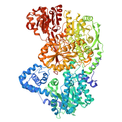
    The proline catabolic pathway consisting of proline dehydrogenase (PRODH) and L-glutamate-γ-semialdehyde (GSAL) dehydrogenase (GSALDH) catalyzes the four-electron oxidation of L-proline to L-glutamate. Chemical probes to these enzymes are of interest for their role in cancer and inherited metabolic disease. Here, we report the results of a crystallographic fragment-screening campaign targeting both enzymes. A unique aspect of our approach is the screening of both enzymes simultaneously using crystals of the bifunctional PRODH-GSALDH enzyme, proline utilization A (PutA). A 288-fragment library from Zenobia was screened in crystallo in cocktails of six fragments. Validation X-ray crystallography with individual fragments identified seven crystal hits distributed in the PRODH active site, GSALDH aldehyde substrate-binding site, and GSALDH NAD + adenine-binding site. The fragment bound in the PRODH active site, 4-methoxybenzyl alcohol, is structurally distinct from all known PRODH inhibitors as it lacks an anionic anchor and stabilizes open conformations of the active site, motivating the study of eighteen analogs. In total, thirteen crystal structures with resolutions ranging from 1.32 Å to 1.80 Å were determined, resolving the poses and interactions of seven fragments from the Zenobia library and five analogs of 4-methoxybenzyl alcohol. These results expand the chemical space of probes targeting proline catabolic enzymes and provide new structural information for further inhibitor development.


  • Organizational Affiliation
    • Department of Biochemistry, University of Missouri, Columbia, MO 65211, USA.

Macromolecule Content 

  • Total Structure Weight: 267.58 kDa 
  • Atom Count: 19,251 
  • Modeled Residue Count: 2,404 
  • Deposited Residue Count: 2,470 
  • Unique protein chains: 1

Macromolecules

Find similar proteins by:|  3D Structure
Entity ID: 1
MoleculeChains  Sequence LengthOrganismDetailsImage
Bifunctional protein PutA
A, B
1,235Sinorhizobium meliloti SM11Mutation(s): 0 
Gene Names: putASM11_chr0102
EC: 1.5.5.2 (PDB Primary Data), 1.2.1.88 (PDB Primary Data)
UniProt
Find proteins for F7X6I3 (Sinorhizobium meliloti (strain SM11))
Explore F7X6I3 
Go to UniProtKB:  F7X6I3
Entity Groups
Sequence Clusters30% Identity50% Identity70% Identity90% Identity95% Identity100% Identity
UniProt GroupF7X6I3
Sequence Annotations
Expand
Reference Sequence

Small Molecules

Ligands 6 Unique
IDChains Name / Formula / InChI Key2D Diagram3D Interactions
FAD
(Subject of Investigation/LOI)

Query on FAD


Download:Ideal Coordinates CCD File
O [auth A],
Y [auth B]
FLAVIN-ADENINE DINUCLEOTIDE
C27 H33 N9 O15 P2
VWWQXMAJTJZDQX-UYBVJOGSSA-N
PGE

Query on PGE


Download:Ideal Coordinates CCD File
C [auth A],
P [auth B]
TRIETHYLENE GLYCOL
C6 H14 O4
ZIBGPFATKBEMQZ-UHFFFAOYSA-N
PYS
(Subject of Investigation/LOI)

Query on PYS


Download:Ideal Coordinates CCD File
E [auth A],
R [auth B]
2-PYRIDINETHIOL
C5 H5 N S
WHMDPDGBKYUEMW-UHFFFAOYSA-N
PEG

Query on PEG


Download:Ideal Coordinates CCD File
D [auth A]
F [auth A]
G [auth A]
H [auth A]
J [auth A]
D [auth A],
F [auth A],
G [auth A],
H [auth A],
J [auth A],
Q [auth B],
S [auth B],
T [auth B]
DI(HYDROXYETHYL)ETHER
C4 H10 O3
MTHSVFCYNBDYFN-UHFFFAOYSA-N
SO4

Query on SO4


Download:Ideal Coordinates CCD File
K [auth A]
L [auth A]
M [auth A]
N [auth A]
W [auth B]
K [auth A],
L [auth A],
M [auth A],
N [auth A],
W [auth B],
X [auth B]
SULFATE ION
O4 S
QAOWNCQODCNURD-UHFFFAOYSA-L
FMT

Query on FMT


Download:Ideal Coordinates CCD File
I [auth A],
U [auth B],
V [auth B]
FORMIC ACID
C H2 O2
BDAGIHXWWSANSR-UHFFFAOYSA-N

Experimental Data & Validation

Experimental Data

  • Method: X-RAY DIFFRACTION
  • Resolution: 1.80 Å
  • R-Value Free:  0.224 (Depositor), 0.227 (DCC) 
  • R-Value Work:  0.194 (Depositor), 0.196 (DCC) 
  • R-Value Observed: 0.196 (Depositor) 
Space Group: P 1 21 1
Unit Cell:
Length ( Å )Angle ( ˚ )
a = 101.155α = 90
b = 102.373β = 106.29
c = 127.504γ = 90
Software Package:
Software NamePurpose
PHENIXrefinement
Aimlessdata scaling
XDSdata reduction
PHASERphasing

Structure Validation

View Full Validation Report


Ligand Structure Quality Assessment 


Entry History 

& Funding Information

Deposition Data


Funding OrganizationLocationGrant Number
National Institutes of Health/National Institute of General Medical Sciences (NIH/NIGMS)United States1R01GM132640

Revision History  (Full details and data files)

  • Version 1.0: 2024-11-27
    Type: Initial release
  • Version 1.1: 2024-12-11
    Changes: Database references