9DCJ | pdb_00009dcj

Pentameric Structure of MERS-CoV Envelope Protein Transmembrane Domain Determined by Solid-State NMR


Experimental Data Snapshot

  • Method: SOLID-STATE NMR
  • Conformers Calculated: 200 
  • Conformers Submitted: 10 
  • Selection Criteria: structures with the lowest energy 

wwPDB Validation 3D Report Full Report

Validation slider image for 9DCJ

This is version 1.1 of the entry. See complete history

Literature

Ion channel structure and function of the MERS coronavirus E protein.

Sucec, I.Xia, B.Somberg, N.H.Wang, Y.Jo, H.Li, S.Perrone, B.Gao, Z.Hong, M.

(2025) Sci Adv 11: eadx1788-eadx1788

  • DOI: https://doi.org/10.1126/sciadv.adx1788
  • Primary Citation Related Structures: 
    9DCJ

  • PubMed Abstract: 

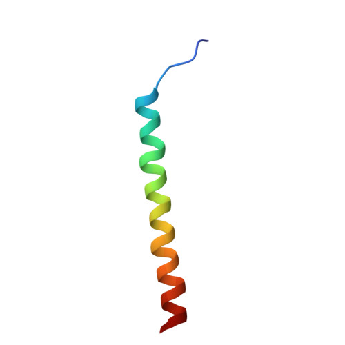
    Coronavirus envelope (E) proteins form drug-targeted ion channels that cause virulence to infected cells. The Middle East respiratory syndrome (MERS) virus has high mortality rates, but its E structure and function are unknown. We report the single-channel conductance and structure of membrane-bound MERS E protein. MERS E conducts K + ions with a unitary conductance of 113 picosiemens, fivefold larger than the conductance of severe acute respiratory syndrome coronavirus 2 E. Solid-state nuclear magnetic resonance data indicate that the MERS E transmembrane domain forms a five-helix bundle that spans the lipid bilayer. The amino-terminal helical interface features multiple interacting phenylalanine (Phe) residues and an asparagine (Asn), whereas the carboxyl-terminal channel pore contains Phe 33 . Mutation of Phe 17 abolished K + conductance, whereas mutations of Phe 33 and Asn 15 suppressed most channel activity. These results indicate that MERS E contains two Phe-centered ion-conduction apparatuses, which likely permeate ions through cation-π interactions, providing the structural basis for developing antiviral drugs to inhibit this pathogenic viroporin.


  • Organizational Affiliation
    • Department of Chemistry, Massachusetts Institute of Technology, 170 Albany Street, Cambridge, MA 02139, USA.

Macromolecule Content 

  • Total Structure Weight: 21.55 kDa 
  • Atom Count: 1,520 
  • Modeled Residue Count: 190 
  • Deposited Residue Count: 190 
  • Unique protein chains: 1

Macromolecules

Find similar proteins by:|  3D Structure
Entity ID: 1
MoleculeChains  Sequence LengthOrganismDetailsImage
Envelope small membrane protein
A, B, C, D, E
38Betacoronavirus England 1Mutation(s): 2 
Gene Names: EsM4
UniProt
Find proteins for K9N5R3 (Middle East respiratory syndrome-related coronavirus (isolate United Kingdom/H123990006/2012))
Explore K9N5R3 
Go to UniProtKB:  K9N5R3
Entity Groups
Sequence Clusters30% Identity50% Identity70% Identity90% Identity95% Identity100% Identity
UniProt GroupK9N5R3
Sequence Annotations
Expand
Reference Sequence

Experimental Data & Validation

Experimental Data

  • Method: SOLID-STATE NMR
  • Conformers Calculated: 200 
  • Conformers Submitted: 10 
  • Selection Criteria: structures with the lowest energy 

Structure Validation

View Full Validation Report


Entry History 

& Funding Information

Deposition Data


Funding OrganizationLocationGrant Number
National Institutes of Health/National Institute of General Medical Sciences (NIH/NIGMS)United States6951834

Revision History  (Full details and data files)

  • Version 1.0: 2025-09-24
    Type: Initial release
  • Version 1.1: 2026-04-08
    Changes: Database references