9DB0 | pdb_00009db0

Molecular basis of pathogenicity of the recently emerged FCoV-23 coronavirus. FCoV-23 S short


Experimental Data Snapshot

  • Method: ELECTRON MICROSCOPY
  • Resolution: 2.50 Å
  • Aggregation State: PARTICLE 
  • Reconstruction Method: SINGLE PARTICLE 

wwPDB Validation 3D Report Full Report

Validation slider image for 9DB0

This is version 1.2 of the entry. See complete history. 

Literature

Loss of FCoV-23 spike domain 0 enhances fusogenicity and entry kinetics.

Tortorici, M.A., Choi, A., Gibson, C.A., Lee, J., Brown, J.T., Stewart, C., Joshi, A., Harari, S., Willoughby, I., Treichel, C., Leaf, E.M., Bloom, J.D., King, N.P., Tait-Burkard, C., Whittaker, G.R., Veesler, D.

(2025) Nature 645: 235-243

  • DOI: https://doi.org/10.1038/s41586-025-09155-z
  • Primary Citation Related Structures: 
    9DAZ, 9DB0, 9DB1, 9DB3, 9DBE, 9DBZ

  • PubMed Abstract: 

    The ability of coronaviruses to recombine and cross species barriers affects human and animal health globally and is a pandemic threat 1,2 . FCoV-23 is a recently emerged, highly pathogenic recombinant coronavirus responsible for a widespread outbreak of feline infectious peritonitis. Here we report cryogenic electron microscopy structures of two FCoV-23 spike isoforms that correspond to the in-host loss of domain 0 observed in clinical samples. The loss of domain 0 markedly enhances the fusogenicity and kinetics of entry into cells and possibly enables biotype switching and lethality. We show that FCoV-23 can use several aminopeptidase N orthologues as receptors and reveal the molecular determinants of receptor species tropism, including a glycan that modulates human receptor engagement. We define antigenic relationships among alphacoronaviruses that infect humans and other mammalian species and identify a cross-reactive alphacoronavirus monoclonal antibody that inhibits FCoV-23 entry. Our results pave the way for the development of vaccines and therapeutics that target this highly pathogenic virus.


  • Organizational Affiliation: 
    • Department of Biochemistry, University of Washington, Seattle, WA, USA.

Macromolecule Content 

  • Total Structure Weight: 440.98 kDa 
  • Atom Count: 24,012 
  • Modeled Residue Count: 2,988 
  • Deposited Residue Count: 3,783 
  • Unique protein chains: 1

Macromolecules

Find similar proteins by:|  3D Structure
Entity ID: 1
MoleculeChains  Sequence LengthOrganismDetailsImage
Spike glycoprotein
A, B, C
1,261Feline coronavirusMutation(s): 0 
Entity Groups
Sequence Clusters30% Identity50% Identity70% Identity90% Identity95% Identity100% Identity
Glycosylation
Glycosylation Sites: 20
Sequence Annotations
Expand
Reference Sequence

Oligosaccharides

Help  
Entity ID: 3
MoleculeChains Length2D Diagram GlycosylationD Interactions
beta-D-mannopyranose-(1-4)-2-acetamido-2-deoxy-beta-D-glucopyranose-(1-4)-2-acetamido-2-deoxy-beta-D-glucopyranose
I,
IA [auth i],
K,
KA [auth k],
V,
I,
IA [auth i],
K,
KA [auth k],
V,
X
3N-Glycosylation
Glycosylation Resources
GlyTouCan: G15407YE
GlyCosmos: G15407YE
GlyGen: G15407YE
Entity ID: 4
MoleculeChains Length2D Diagram GlycosylationD Interactions
alpha-D-mannopyranose-(1-3)-[alpha-D-mannopyranose-(1-6)]beta-D-mannopyranose-(1-4)-2-acetamido-2-deoxy-beta-D-glucopyranose-(1-4)-2-acetamido-2-deoxy-beta-D-glucopyranoseL,
LA [auth l],
Y
5N-Glycosylation
Glycosylation Resources
GlyTouCan: G22768VO
GlyCosmos: G22768VO
GlyGen: G22768VO
Entity ID: 5
MoleculeChains Length2D Diagram GlycosylationD Interactions
alpha-D-mannopyranose-(1-3)-beta-D-mannopyranose-(1-4)-2-acetamido-2-deoxy-beta-D-glucopyranose-(1-4)-2-acetamido-2-deoxy-beta-D-glucopyranoseAA [auth a],
N,
NA [auth n]
4N-Glycosylation
Glycosylation Resources
GlyTouCan: G81315DD
GlyCosmos: G81315DD
GlyGen: G81315DD

Small Molecules

Ligands 2 Unique
IDChains Name / Formula / InChI Key2D Diagram3D Interactions
PAM
(Subject of Investigation/LOI)

Query on PAM


Download:Ideal Coordinates CCD File
GB [auth B]
IB [auth C]
PB [auth C]
WA [auth A]
YA [auth A]
GB [auth B],
IB [auth C],
PB [auth C],
WA [auth A],
YA [auth A],
ZA [auth B]
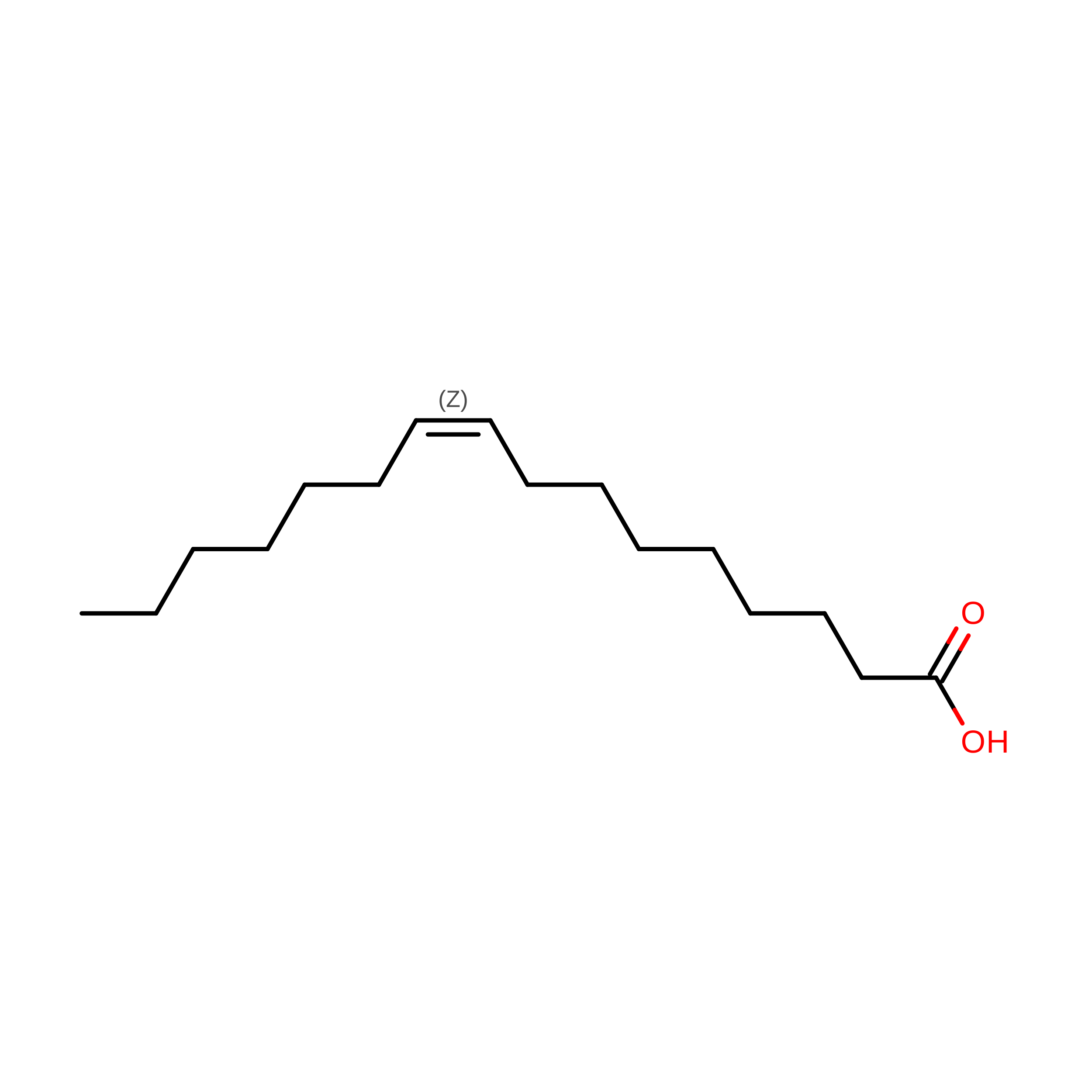
PALMITOLEIC ACID
C16 H30 O2
SECPZKHBENQXJG-FPLPWBNLSA-N
NAG
(Subject of Investigation/LOI)

Query on NAG


Download:Ideal Coordinates CCD File
AB [auth B]
BB [auth B]
CB [auth B]
DB [auth B]
EB [auth B]
AB [auth B],
BB [auth B],
CB [auth B],
DB [auth B],
EB [auth B],
FB [auth B],
HB [auth B],
JB [auth C],
KB [auth C],
LB [auth C],
MB [auth C],
NB [auth C],
OB [auth C],
QA [auth A],
QB [auth C],
RA [auth A],
SA [auth A],
TA [auth A],
UA [auth A],
VA [auth A],
XA [auth A]
2-acetamido-2-deoxy-beta-D-glucopyranose
C8 H15 N O6
OVRNDRQMDRJTHS-FMDGEEDCSA-N

Experimental Data & Validation

Experimental Data

  • Method: ELECTRON MICROSCOPY
  • Resolution: 2.50 Å
  • Aggregation State: PARTICLE 
  • Reconstruction Method: SINGLE PARTICLE 

Structure Validation

View Full Validation Report


Entry History 

& Funding Information

Deposition Data


Funding OrganizationLocationGrant Number
National Institutes of Health/National Institute Of Allergy and Infectious Diseases (NIH/NIAID)United StatesAI158186

Revision History  (Full details and data files)

  • Version 1.0: 2025-07-09
    Type: Initial release
  • Version 1.1: 2025-07-23
    Changes: Data collection, Database references
  • Version 1.2: 2025-09-17
    Changes: Data collection, Database references