9D59 | pdb_00009d59

Structure of Citrobacter multi-ubiquitin protein filament


Experimental Data Snapshot

  • Method: ELECTRON MICROSCOPY
  • Resolution: 2.43 Å
  • Aggregation State: FILAMENT 
  • Reconstruction Method: HELICAL 

wwPDB Validation 3D Report Full Report

Validation slider image for 9D59

This is version 1.2 of the entry. See complete history

Literature

Structural diversity and oligomerization of bacterial ubiquitin-like proteins.

Gong, M.Ye, Q.Gu, Y.Chambers, L.R.Bobkov, A.A.Arakawa, N.K.Matyszewski, M.Corbett, K.D.

(2025) Structure 33: 1016-1026.e4

  • DOI: https://doi.org/10.1016/j.str.2025.03.011
  • Primary Citation Related Structures: 
    8U38, 9CD2, 9D59, 9D5A, 9D5B

  • PubMed Abstract: 

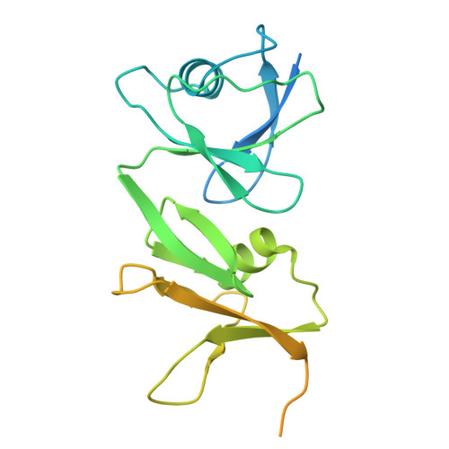
    Bacteria possess a variety of operons with homology to eukaryotic ubiquitination pathways that encode predicted E1, E2, E3, deubiquitinase, and ubiquitin-like proteins. Some of these pathways have recently been shown to function in anti-bacteriophage immunity, but the biological functions of others remain unknown. Here, we show that ubiquitin-like proteins in two bacterial operon families show surprising architectural diversity, possessing one to three β-grasp domains preceded by diverse N-terminal domains. We find that a large group of bacterial ubiquitin-like proteins possess three β-grasp domains and form homodimers and helical filaments mediated by conserved Ca 2+ ion binding sites. Our findings highlight a distinctive mode of self-assembly for ubiquitin-like proteins and suggest that Ca 2+ -mediated ubiquitin-like protein filament assembly and/or disassembly enables cells to sense and respond to stress conditions that alter intracellular metal ion concentration.


  • Organizational Affiliation
    • Department of Cellular and Molecular Medicine, University of California, San Diego, La Jolla, CA 92093, USA.

Macromolecule Content 

  • Total Structure Weight: 114.07 kDa 
  • Atom Count: 4,698 
  • Modeled Residue Count: 584 
  • Deposited Residue Count: 988 
  • Unique protein chains: 1

Macromolecules

Find similar proteins by:|  3D Structure
Entity ID: 1
MoleculeChains  Sequence LengthOrganismDetailsImage
Multi-ubiquitin domain-containing protein
A, B, C, D
247Citrobacter sp. RHBSTW-00271Mutation(s): 0 
UniProt
Find proteins for A0A5B0SRH6 (Citrobacter portucalensis)
Explore A0A5B0SRH6 
Go to UniProtKB:  A0A5B0SRH6
Entity Groups
Sequence Clusters30% Identity50% Identity70% Identity90% Identity95% Identity100% Identity
UniProt GroupA0A5B0SRH6
Sequence Annotations
Expand
Reference Sequence

Small Molecules

Ligands 1 Unique
IDChains Name / Formula / InChI Key2D Diagram3D Interactions
CA
(Subject of Investigation/LOI)

Query on CA


Download:Ideal Coordinates CCD File
E [auth A]
F [auth A]
G [auth A]
H [auth A]
I [auth B]
E [auth A],
F [auth A],
G [auth A],
H [auth A],
I [auth B],
J [auth B],
K [auth B],
L [auth B],
M [auth C],
N [auth C],
O [auth C],
P [auth D],
Q [auth D],
R [auth D]
CALCIUM ION
Ca
BHPQYMZQTOCNFJ-UHFFFAOYSA-N

Experimental Data & Validation

Experimental Data

  • Method: ELECTRON MICROSCOPY
  • Resolution: 2.43 Å
  • Aggregation State: FILAMENT 
  • Reconstruction Method: HELICAL 
EM Software:
TaskSoftware PackageVersion
MODEL REFINEMENTPHENIX1.21.1_5286

Structure Validation

View Full Validation Report


Entry History 

& Funding Information

Deposition Data


Funding OrganizationLocationGrant Number
National Institutes of Health/National Institute of General Medical Sciences (NIH/NIGMS)United StatesR35 GM144121

Revision History  (Full details and data files)

  • Version 1.0: 2025-04-02
    Type: Initial release
  • Version 1.1: 2025-04-30
    Changes: Data collection, Database references
  • Version 1.2: 2025-06-18
    Changes: Data collection, Database references