9BOU | pdb_00009bou

Crystal structure of ATP-grasp ligase PruB from Streptomyces coelicolor A3(2)


Experimental Data Snapshot

  • Method: X-RAY DIFFRACTION
  • Resolution: 2.91 Å
  • R-Value Free: 
    0.258 (Depositor), 0.262 (DCC) 
  • R-Value Work: 
    0.207 (Depositor), 0.209 (DCC) 
  • R-Value Observed: 
    0.210 (Depositor) 

Starting Model: experimental
View more details

wwPDB Validation   3D Report Full Report


This is version 1.1 of the entry. See complete history


Literature

Characterization of a Dual Function Peptide Cyclase in Graspetide Biosynthesis.

Rubin, G.M.Patel, K.P.Jiang, Y.Ishee, A.C.Seabra, G.Bruner, S.D.Ding, Y.

(2024) ACS Chem Biol 19: 2525-2534

  • DOI: https://doi.org/10.1021/acschembio.4c00626
  • Primary Citation of Related Structures:  
    9BOU

  • PubMed Abstract: 

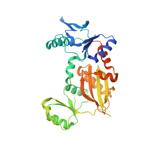
    Graspetides are a diverse family of ribosomally synthesized and post-translationally modified peptides with unique macrocyclic structures formed by ATP-grasp enzymes. Group 11 graspetides, including prunipeptin, feature both macrolactone and macrolactam cross-links. Despite the known involvement of a single ATP-grasp cyclase in the dual macrocyclizations of groups 5, 7, and 11 graspetides, detailed mechanistic insights into these enzymes remain limited. Here, we reconstructed prunipeptin biosynthesis from Streptomyces coelicolor using recombinant PruA and PruB macrocyclase. PruB exhibited kinetic behavior similar to other characterized graspetide cyclases, with a notably higher k cat , likely due to utilization of an ATP-regeneration system. The X-ray crystal structure of PruB revealed distinct features as compared to groups 1 and 2 enzymes. Site-directed mutagenesis identified critical roles of key residues for the PruB reaction, including the DxR motif conserved in other graspetide cyclases. Additionally, computational modeling of the PruA/PruB cocomplex uncovered substrate interactions and suggested that PruB first catalyzes a macrolactone bond formation on PruA. This study enhances our understanding of ATP-grasp enzyme mechanisms in graspetide biosynthesis and provides insights for engineering these enzymes for future applications.


  • Organizational Affiliation
    • Department of Medicinal Chemistry, Center for Natural Products, Drug Discovery and Development, University of Florida, Gainesville, Florida 31610, United States.

Macromolecules
Find similar proteins by:  (by identity cutoff)  |  3D Structure
Entity ID: 1
MoleculeChains Sequence LengthOrganismDetailsImage
MvdD-like pre-ATP grasp domain-containing protein
A, B
339Streptomyces coelicolor A3(2)Mutation(s): 0 
Gene Names: SCO0695SCF42.05
UniProt
Find proteins for Q9L2K8 (Streptomyces coelicolor (strain ATCC BAA-471 / A3(2) / M145))
Explore Q9L2K8 
Go to UniProtKB:  Q9L2K8
Entity Groups  
Sequence Clusters30% Identity50% Identity70% Identity90% Identity95% Identity100% Identity
UniProt GroupQ9L2K8
Sequence Annotations
Expand
  • Reference Sequence
Experimental Data & Validation

Experimental Data

  • Method: X-RAY DIFFRACTION
  • Resolution: 2.91 Å
  • R-Value Free:  0.258 (Depositor), 0.262 (DCC) 
  • R-Value Work:  0.207 (Depositor), 0.209 (DCC) 
  • R-Value Observed: 0.210 (Depositor) 
Space Group: I 4 2 2
Unit Cell:
Length ( Å )Angle ( ˚ )
a = 222.04α = 90
b = 222.04β = 90
c = 87.007γ = 90
Software Package:
Software NamePurpose
PHENIXrefinement
XDSdata reduction
Aimlessdata scaling
PHASERphasing

Structure Validation

View Full Validation Report


Entry History & Funding Information

Deposition Data


Funding OrganizationLocationGrant Number
National Institutes of Health/National Institute of General Medical Sciences (NIH/NIGMS)United StatesR35GM128742
National Institutes of Health/National Institute of General Medical Sciences (NIH/NIGMS)United StatesGM145426

Revision History  (Full details and data files)

  • Version 1.0: 2024-12-18
    Type: Initial release
  • Version 1.1: 2025-01-01
    Changes: Database references