9BF6 | pdb_00009bf6

Cryo-EM structure of the HIV-1 WITO IDL Env trimer in complex with PGT122 Fab


Experimental Data Snapshot

  • Method: ELECTRON MICROSCOPY
  • Resolution: 4.50 Å
  • Aggregation State: PARTICLE 
  • Reconstruction Method: SINGLE PARTICLE 

Starting Model: in silico
View more details

wwPDB Validation 3D Report Full Report

Validation slider image for 9BF6

This is version 1.1 of the entry. See complete history

Literature

Design of soluble HIV-1 envelope trimers free of covalent gp120-gp41 bonds with prevalent native-like conformation.

Zhang, P.Gorman, J.Tsybovsky, Y.Lu, M.Liu, Q.Gopan, V.Singh, M.Lin, Y.Miao, H.Seo, Y.Kwon, A.Olia, A.S.Chuang, G.Y.Geng, H.Lai, Y.T.Zhou, T.Mascola, J.R.Mothes, W.Kwong, P.D.Lusso, P.

(2024) Cell Rep 43: 114518-114518

  • DOI: https://doi.org/10.1016/j.celrep.2024.114518
  • Primary Citation Related Structures: 
    9BER, 9BEW, 9BF6

  • PubMed Abstract: 

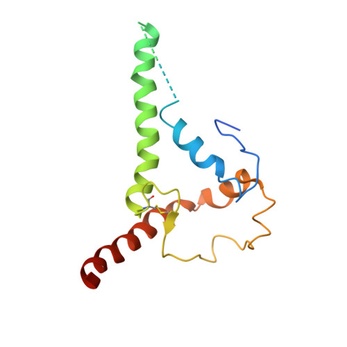
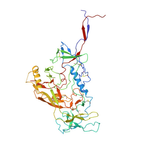
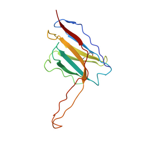
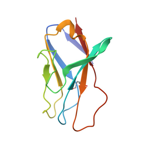
    Soluble HIV-1 envelope (Env) trimers may serve as effective vaccine immunogens. The widely utilized SOSIP trimers have been paramount for structural studies, but the disulfide bond they feature between gp120 and gp41 constrains intersubunit mobility and may alter antigenicity. Here, we report an alternative strategy to generate stabilized soluble Env trimers free of covalent gp120-gp41 bonds. Stabilization was achieved by introducing an intrasubunit disulfide bond between the inner and outer domains of gp120, defined as interdomain lock (IDL). Correctly folded IDL trimers displaying a native-like antigenic profile were produced for HIV-1 Envs of different clades. Importantly, the IDL design abrogated CD4 binding while not affecting recognition by potent neutralizing antibodies to the CD4-binding site. By cryoelectron microscopy, IDL trimers were shown to adopt a closed prefusion configuration, while single-molecule fluorescence resonance energy transfer documented a high prevalence of native-like conformation. Thus, IDL trimers may be promising candidates as vaccine immunogens.


  • Organizational Affiliation
    • Laboratory of Immunoregulation, National Institute of Allergy and Infectious Diseases, National Institutes of Health, Bethesda, MD 20892, USA. Electronic address: zhangp@ihm.ac.cn.

Macromolecule Content 

  • Total Structure Weight: 318.19 kDa 
  • Atom Count: 20,330 
  • Modeled Residue Count: 2,367 
  • Deposited Residue Count: 2,610 
  • Unique protein chains: 4

Macromolecules

Find similar proteins by:|  3D Structure
Entity ID: 1
MoleculeChains  Sequence LengthOrganismDetailsImage
Envelope glycoprotein gp41A,
E [auth B],
I
153Human immunodeficiency virus 1Mutation(s): 0 
Gene Names: env
UniProt
Find proteins for Q5G5U5 (Human immunodeficiency virus type 1)
Explore Q5G5U5 
Go to UniProtKB:  Q5G5U5
Entity Groups
Sequence Clusters30% Identity50% Identity70% Identity90% Identity95% Identity100% Identity
UniProt GroupQ5G5U5
Glycosylation
Glycosylation Sites: 4
Sequence Annotations
Expand
Reference Sequence
Find similar proteins by:|  3D Structure
Entity ID: 2
MoleculeChains  Sequence LengthOrganismDetailsImage
Envelope glycoprotein gp120B [auth C],
F,
J
480Human immunodeficiency virus 1Mutation(s): 0 
Gene Names: env
UniProt
Find proteins for Q5G5U5 (Human immunodeficiency virus type 1)
Explore Q5G5U5 
Go to UniProtKB:  Q5G5U5
Entity Groups
Sequence Clusters30% Identity50% Identity70% Identity90% Identity95% Identity100% Identity
UniProt GroupQ5G5U5
Glycosylation
Glycosylation Sites: 18
Sequence Annotations
Expand
Reference Sequence
Find similar proteins by:|  3D Structure
Entity ID: 3
MoleculeChains  Sequence LengthOrganismDetailsImage
PGT122 heavy chainC [auth D],
G,
K
132Homo sapiensMutation(s): 0 
Find similar proteins by:|  3D Structure
Entity ID: 4
MoleculeChains  Sequence LengthOrganismDetailsImage
PGT122 light chainD [auth E],
H,
L
105Homo sapiensMutation(s): 0 

Oligosaccharides

Help  
Entity ID: 5
MoleculeChains Length2D Diagram GlycosylationD Interactions
beta-D-mannopyranose-(1-4)-2-acetamido-2-deoxy-beta-D-glucopyranose-(1-4)-2-acetamido-2-deoxy-beta-D-glucopyranose
AA [auth a],
CA [auth c],
EA [auth e],
GA [auth g],
M,
AA [auth a],
CA [auth c],
EA [auth e],
GA [auth g],
M,
O,
Q,
S,
T,
V,
X,
Z
3N-Glycosylation
Glycosylation Resources
GlyTouCan: G15407YE
GlyCosmos: G15407YE
GlyGen: G15407YE
Entity ID: 6
MoleculeChains Length2D Diagram GlycosylationD Interactions
alpha-D-mannopyranose-(1-2)-alpha-D-mannopyranose-(1-3)-[alpha-D-mannopyranose-(1-6)]beta-D-mannopyranose-(1-4)-2-acetamido-2-deoxy-beta-D-glucopyranose-(1-4)-2-acetamido-2-deoxy-beta-D-glucopyranose
BA [auth b],
DA [auth d],
N,
P,
U,
BA [auth b],
DA [auth d],
N,
P,
U,
W
6N-Glycosylation
Glycosylation Resources
GlyTouCan: G56014GC
GlyCosmos: G56014GC
GlyGen: G56014GC
Entity ID: 7
MoleculeChains Length2D Diagram GlycosylationD Interactions
alpha-D-mannopyranose-(1-3)-beta-D-mannopyranose-(1-4)-2-acetamido-2-deoxy-beta-D-glucopyranose-(1-4)-2-acetamido-2-deoxy-beta-D-glucopyranoseFA [auth f],
R,
Y
4N-Glycosylation
Glycosylation Resources
GlyTouCan: G81315DD
GlyCosmos: G81315DD
GlyGen: G81315DD

Small Molecules

Ligands 2 Unique
IDChains Name / Formula / InChI Key2D Diagram3D Interactions
NAG

Query on NAG


Download:Ideal Coordinates CCD File
AB [auth F]
AC [auth J]
BB [auth F]
CB [auth F]
DB [auth F]
AB [auth F],
AC [auth J],
BB [auth F],
CB [auth F],
DB [auth F],
EB [auth F],
FB [auth F],
GB [auth F],
HA [auth A],
HB [auth F],
IA [auth A],
IB [auth F],
JA [auth A],
JB [auth F],
KA [auth A],
KB [auth F],
LA [auth C],
MA [auth C],
MB [auth I],
NA [auth C],
NB [auth I],
OA [auth C],
OB [auth I],
PA [auth C],
PB [auth I],
QA [auth C],
QB [auth J],
RA [auth C],
RB [auth J],
SA [auth C],
SB [auth J],
TA [auth C],
TB [auth J],
UA [auth C],
UB [auth J],
VA [auth C],
VB [auth J],
WA [auth B],
WB [auth J],
XA [auth B],
XB [auth J],
YA [auth B],
YB [auth J],
ZA [auth B],
ZB [auth J]
2-acetamido-2-deoxy-beta-D-glucopyranose
C8 H15 N O6
OVRNDRQMDRJTHS-FMDGEEDCSA-N
PO4

Query on PO4


Download:Ideal Coordinates CCD File
LB [auth F]PHOSPHATE ION
O4 P
NBIIXXVUZAFLBC-UHFFFAOYSA-K

Experimental Data & Validation

Experimental Data

  • Method: ELECTRON MICROSCOPY
  • Resolution: 4.50 Å
  • Aggregation State: PARTICLE 
  • Reconstruction Method: SINGLE PARTICLE 
EM Software:
TaskSoftware PackageVersion
RECONSTRUCTIONcryoSPARC2.14
MODEL REFINEMENTPHENIX

Structure Validation

View Full Validation Report


Entry History 

& Funding Information

Deposition Data


Funding OrganizationLocationGrant Number
National Institutes of Health/National Institute of General Medical Sciences (NIH/NIGMS)United StatesGM103310
Simons FoundationUnited StatesSF349247

Revision History  (Full details and data files)

  • Version 1.0: 2024-07-31
    Type: Initial release
  • Version 1.1: 2024-10-23
    Changes: Data collection, Structure summary