9AZ6 | pdb_00009az6

F-actin-Talin(R13-DD) complex


Experimental Data Snapshot

  • Method: ELECTRON MICROSCOPY
  • Resolution: 2.98 Å
  • Aggregation State: FILAMENT 
  • Reconstruction Method: HELICAL 

Starting Models: experimental
View more details

wwPDB Validation   3D Report Full Report


This is version 1.0 of the entry. See complete history


Literature

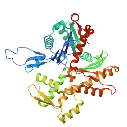
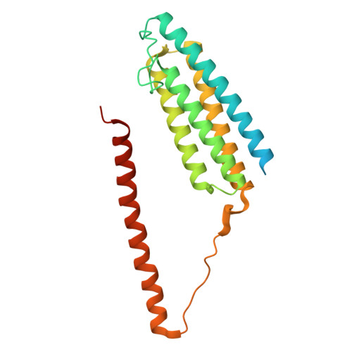
Biochemical and structural bases for talin ABSs-F-actin interactions.

Biertumpfel, C.Yamada, Y.Vasquez-Montes, V.Truong, T.V.Cada, A.K.Mizuno, N.

(2025) Proc Natl Acad Sci U S A 122: e2405922122-e2405922122

  • DOI: https://doi.org/10.1073/pnas.2405922122
  • Primary Citation of Related Structures:  
    9AZ6

  • PubMed Abstract: 

    Focal adhesions (FAs) are large intracellular macromolecular assemblies that play a critical role in cell polarization and migration. Talin serves as a direct connection between integrin receptor and actomyosin cytoskeleton within FAs. Talin contains three actin-binding sites (ABS1-3) that engage discreetly during the development of FAs, thus acting as a critical player in FA initiation and maturation. However, the molecular basis of the ABS-F-actin interactions remains unknown. Here, we explore interactions of ABSs with F-actin to understand the multivalent behavior of talin. Particularly, the cryo-EM structure of the F-actin-ABS3 complex at 2.9 Å shows ABS3 spanning through two actin monomers along the filament axis, each occupied by the R13 rod subdomain and the DD domain. The dimerization of ABS3 occurs through the DD domain where both protomers interact on the actin surface, and the dimerization of talin to the actin surface is necessary for the engagement to F-actin. The R13 helical bundle is distorted upon binding to F-actin and releases the H1 helix from the rest of the bundle. This phenomenon has also been observed with other tension-sensing proteins like vinculin and α-catenin, highlighting that unfolding is relevant for its force sensing activity. On the contrary, ABS2 (R4R8 subdomains), which is thought to be critical for the maintenance of mature FAs, had multiple F-actin-binding regions within ABS2 and the binding likely occurred by these subdomains running through the surface of F-actin, thus strengthening the interactions upon the maturation of FAs.


  • Organizational Affiliation
    • Laboratory of Structural Cell Biology, National Heart, Lung, and Blood Institute, NIH, Bethesda, MD 20892.

Macromolecules
Find similar proteins by:  (by identity cutoff)  |  3D Structure
Entity ID: 1
MoleculeChains Sequence LengthOrganismDetailsImage
Actin, alpha skeletal muscle
A, B, C, D, E
375Oryctolagus cuniculusMutation(s): 0 
EC: 3.6.4
UniProt
Find proteins for P68135 (Oryctolagus cuniculus)
Explore P68135 
Go to UniProtKB:  P68135
Entity Groups  
Sequence Clusters30% Identity50% Identity70% Identity90% Identity95% Identity100% Identity
UniProt GroupP68135
Sequence Annotations
Expand
  • Reference Sequence
Find similar proteins by:  (by identity cutoff)  |  3D Structure
Entity ID: 2
MoleculeChains Sequence LengthOrganismDetailsImage
Talin-1
F, G, H, I, J
F, G, H, I, J, K, L, M
246Mus musculusMutation(s): 0 
Gene Names: Tln1Tln
UniProt
Find proteins for P26039 (Mus musculus)
Explore P26039 
Go to UniProtKB:  P26039
Entity Groups  
Sequence Clusters30% Identity50% Identity70% Identity90% Identity95% Identity100% Identity
UniProt GroupP26039
Sequence Annotations
Expand
  • Reference Sequence
Small Molecules
Ligands 2 Unique
IDChains Name / Formula / InChI Key2D Diagram3D Interactions
ADP (Subject of Investigation/LOI)
Query on ADP

Download Ideal Coordinates CCD File 
N [auth A],
P [auth B],
R [auth C],
T [auth D],
V [auth E]
ADENOSINE-5'-DIPHOSPHATE
C10 H15 N5 O10 P2
XTWYTFMLZFPYCI-KQYNXXCUSA-N
MG (Subject of Investigation/LOI)
Query on MG

Download Ideal Coordinates CCD File 
O [auth A],
Q [auth B],
S [auth C],
U [auth D],
W [auth E]
MAGNESIUM ION
Mg
JLVVSXFLKOJNIY-UHFFFAOYSA-N
Modified Residues  1 Unique
IDChains TypeFormula2D DiagramParent
HIC
Query on HIC
A, B, C, D, E
L-PEPTIDE LINKINGC7 H11 N3 O2HIS
Experimental Data & Validation

Experimental Data

  • Method: ELECTRON MICROSCOPY
  • Resolution: 2.98 Å
  • Aggregation State: FILAMENT 
  • Reconstruction Method: HELICAL 
EM Software:
TaskSoftware PackageVersion
RECONSTRUCTIONRELION3.1.0
MODEL REFINEMENTPHENIX1.21-5207

Structure Validation

View Full Validation Report


Entry History & Funding Information

Deposition Data


Funding OrganizationLocationGrant Number
National Institutes of Health/National Heart, Lung, and Blood Institute (NIH/NHLBI)United States--
National Institutes of Health/National Institute of Arthritis and Musculoskeletal and Skin Diseases (NIH/NIAMS)United States--

Revision History  (Full details and data files)

  • Version 1.0: 2025-02-12
    Type: Initial release