Structural and Biochemical Insights into Lignin-Oxidizing Activity of Bacterial Peroxidases against Soluble Substrates and Kraft Lignin.
Choolaei, Z., Khusnutdinova, A.N., Skarina, T., Stogios, P., Diep, P., Lemak, S., Edwards, E.A., Savchenko, A., Yakunin, A.F.(2025) ACS Chem Biol 20: 830-844
- PubMed: 40145573 Search on PubMed
- DOI: https://doi.org/10.1021/acschembio.4c00788
- Primary Citation Related Structures:
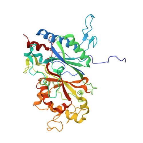
9AZ0, 9AZ1, 9AZ2 - PubMed Abstract:
Great interest has recently been drawn to the production of value-added products from lignin; however, its recalcitrance and high chemical complexity have made this challenging. Dye-decolorizing peroxidases and catalase-peroxidases are among the enzymes that are recognized to play important roles in environmental lignin oxidation. However, bacterial lignin-oxidizing enzymes remain less characterized compared to related proteins from fungi. In this study, screening of 18 purified bacterial peroxidases against the general chromogenic substrate 2,2'-azinobis(3-ethylbenzthiazoline-6-sulfonate) (ABTS) revealed the presence of peroxidase activity in all proteins. Agarose plate-based screens with kraft lignin identified detectable and high lignin oxidation activity in 15 purified proteins. Crystal structures were determined for the DyP-type peroxidases FC2591 from Frankia casuarinae , PF3257 from Pseudomonas fluorescens , and PR9465 from Pseudomonas rhizosphaerae . The structures revealed the presence of hemes with bound oxygens coordinated by conserved His, Arg, and Asp residues as well as three molecular tunnels connecting the heme with the protein surface. Structure-based site-directed mutagenesis of FC2591 identified at least five active site residues as essential for oxidase activity against both ABTS and lignin, whereas the S370A mutant protein showed a three- to 4-fold activity increase with both substrates. HPLC analysis of reaction products of the wild-type FC2591 and S370A mutant proteins with the model lignin dimer guaiacylglycerol-β-guaiacyl ether and kraft lignin revealed the formation of products consistent with the radical coupling of the reaction intermediates. Thus, this study identified novel bacterial heme peroxidases with lignin oxidation activity and provided further insights into our understanding of these enzymes.
- Department of Chemical Engineering and Applied Chemistry, University of Toronto, Toronto M5S 3E5, Canada.
Organizational Affiliation: