


Crystal Structure of a Ribokinase from Brucella suis in complex with ADP
Seibold, S., Lovell, S., Battaile, K.P.To be published.
Experimental Data Snapshot
Starting Model: in silico
View more details
Entity ID: 1 | |||||
|---|---|---|---|---|---|
| Molecule | Chains | Sequence Length | Organism | Details | Image |
| Ribokinase | 312 | Brucella suis 1330 | Mutation(s): 0 Gene Names: rbsK, BS1330_II0005 EC: 2.7.1.15 | 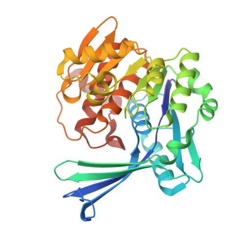 | |
UniProt | |||||
Find proteins for A0A0H3GDY9 (Brucella suis biovar 1 (strain 1330)) Explore A0A0H3GDY9 Go to UniProtKB: A0A0H3GDY9 | |||||
Entity Groups | |||||
| Sequence Clusters | 30% Identity50% Identity70% Identity90% Identity95% Identity100% Identity | ||||
| UniProt Group | A0A0H3GDY9 | ||||
Sequence AnnotationsExpand | |||||
Reference Sequence | |||||
| Ligands 3 Unique | |||||
|---|---|---|---|---|---|
| ID | Chains | Name / Formula / InChI Key | 2D Diagram | 3D Interactions | |
| ADP (Subject of Investigation/LOI) Download:Ideal Coordinates CCD File | BA [auth C] CB [auth F] G [auth A] LA [auth D] Q [auth B] | ADENOSINE-5'-DIPHOSPHATE C10 H15 N5 O10 P2 XTWYTFMLZFPYCI-KQYNXXCUSA-N |  | ||
| SO4 Download:Ideal Coordinates CCD File | CA [auth C] DA [auth C] DB [auth F] EA [auth C] EB [auth F] | SULFATE ION O4 S QAOWNCQODCNURD-UHFFFAOYSA-L |  | ||
| CL Download:Ideal Coordinates CCD File | AA [auth C] AB [auth F] BB [auth F] JA [auth D] KA [auth D] | CHLORIDE ION Cl VEXZGXHMUGYJMC-UHFFFAOYSA-M |  | ||
| Length ( Å ) | Angle ( ˚ ) |
|---|---|
| a = 225.093 | α = 90 |
| b = 138.228 | β = 107.04 |
| c = 80.872 | γ = 90 |
| Software Name | Purpose |
|---|---|
| PHENIX | refinement |
| Aimless | data scaling |
| XDS | data reduction |
| PHASER | phasing |
| PDB_EXTRACT | data extraction |
| Funding Organization | Location | Grant Number |
|---|---|---|
| National Institutes of Health/National Institute Of Allergy and Infectious Diseases (NIH/NIAID) | United States | 75N93022C00036 |
| National Institutes of Health/Office of the Director | United States | S10OD030394 |


