Covalent Binding of the Boronic Acid-Based Inhibitor GSK4394835A to Phosphodiesterase 3B, a Drug Target for Cardiovascular Disease.
Eaton, S.A., Christianson, D.W.(2025) J Med Chem 68: 26574-26578
- PubMed: 41331906 Search on PubMed
- DOI: https://doi.org/10.1021/acs.jmedchem.5c03081
- Primary Citation Related Structures:
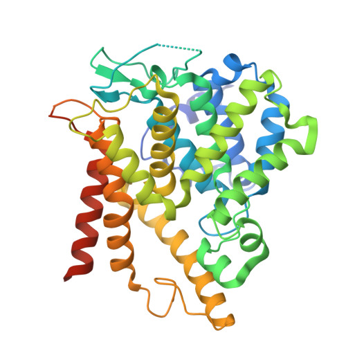
9YUD - PubMed Abstract:
Boronic acid inhibitors often undergo nucleophilic addition upon binding to an enzyme due to the electrophilicity of the boron atom. A new class of boronic acid inhibitors of human phosphodiesterase 3B (PDE3B) has recently been disclosed, along with the 2.7 Å-resolution crystal structure of PDE3B complexed with inhibitor GSK4394835A [Rowley et al. (2024) Discovery and SAR study of boronic acid-based selective PDE3B inhibitors from a novel DNA-encoded library. J. Med. Chem. 67 , 2049-2065]. The crystal structure shows the binding of an intact, unreacted boronic acid, but discrepancies were evident in refinement statistics. Accordingly, we redetermined the structure using structure factor amplitudes deposited in the Protein Data Bank (accession code 8SYC), showing that the boronic acid moiety of GSK4394835A undergoes nucleophilic attack by H737 to form a tetrahedral boronate anion. We refined the structure to convergence with excellent refinement statistics, concluding that GSK4394835A is a reversible covalent inhibitor of PDE3B.
- Roy and Diana Vagelos Laboratories, Department of Chemistry, University of Pennsylvania, Philadelphia, Pennsylvania 19104-6323, United States.
Organizational Affiliation: