9YNZ | pdb_00009ynz

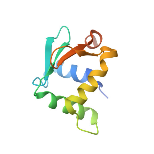
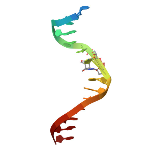
Human PU.1 ETS-Domain (165-270) Bound to d(5'-AATAAGCGGAAGTGGG-3') d(5'-TCCCACT*CPD*CGCTTAT-3')

  • Classification: TRANSCRIPTION
  • Organism(s): synthetic construct, Homo sapiens
  • Expression System: Escherichia coli
  • Mutation(s): No 

  • Deposited: 2025-10-13 Released: 2025-10-22 
  • Deposition Author(s): Terrell, J.R., Poon, G.M.K.
  • Funding Organization(s): National Institutes of Health/National Institute of Environmental Health Sciences (NIH/NIEHS), National Institutes of Health/National Heart, Lung, and Blood Institute (NIH/NHLBI), National Science Foundation (NSF, United States)

Experimental Data Snapshot

  • Method: X-RAY DIFFRACTION
  • Resolution: 2.05 Å
  • R-Value Free: 
    0.244 (Depositor), 0.242 (DCC) 
  • R-Value Work: 
    0.206 (Depositor), 0.206 (DCC) 
  • R-Value Observed: 
    0.210 (Depositor) 

Starting Model: experimental
View more details

wwPDB Validation 3D Report Full Report

Validation slider image for 9YNZ

This is version 1.1 of the entry. See complete history. 

Literature

Molecular basis of UV lesion binding and repair inhibition by ETS-family transcription factors.

Sivapragasam, S., Terrell, J.R., van der Vaart, A., Germann, M.W., Laughery, M.F., Everly, M.E., Adhikari, S.P., Hrdlicka, P.J., Wyrick, J.J., Poon, G.M.K.

(2026) Nucleic Acids Res 54

  • DOI: https://doi.org/10.1093/nar/gkag711
  • Primary Citation Related Structures: 
    9YNZ

  • PubMed Abstract: 

    Mutation hotspots in melanoma frequently occur at DNA binding sites of E26 transformation-specific (ETS)-family transcription factors, as ETS factors stimulate the formation of UV-induced cyclobutane pyrimidine dimers (CPDs) while suppressing repair at ETS-bound DNA sites. To elucidate the molecular mechanism by which ETS factors bind to damaged DNA sites and inhibit repair, we investigated the binding of members from the three major classes of the ETS superfamily (Ets1, ELF1, and PU.1) to cognate DNA containing a cis-syn TpT CPD. These site-specific CPDs modulated ETS recognition and repair by a model repair enzyme in a position-dependent manner. Specifically, a deaminated CPD located in a damage hotspot in the ETS binding motif consistently stimulated binding and inhibited T4 PDG (a CPD repair enzyme) by all three paralogs. Co-crystal structures of PU.1 reveal that CPDs and mismatches are recognized within the framework of canonical ETS/DNA complexes. Molecular dynamics simulations in explicit solvent show that CPD introduces compensatory structural dynamics to both the free and ETS-bound states that strongly modify the underlying thermodynamics of recognition. The results offer a molecular basis for how ETS factors induce mutation hotspots in skin cancers and other UV-exposed tissues by binding to CPD-containing sites and inhibiting their repair.


  • Organizational Affiliation: 
    • School of Molecular Biosciences, Washington State University, Pullman WA 99164,USA.

Macromolecule Content 

  • Total Structure Weight: 22.25 kDa 
  • Atom Count: 1,445 
  • Modeled Residue Count: 121 
  • Deposited Residue Count: 137 
  • Unique protein chains: 1
  • Unique nucleic acid chains: 2

Macromolecules


Find similar proteins by:|  3D Structure
Entity ID: 3
MoleculeChains  Sequence LengthOrganismDetailsImage
Transcription factor PU.1C [auth F]106Homo sapiensMutation(s): 0 
Gene Names: SPI1
UniProt & NIH Common Fund Data Resources
Find proteins for P17947 (Homo sapiens)
Explore P17947 
Go to UniProtKB:  P17947
PHAROS:  P17947
GTEx:  ENSG00000066336 
Entity Groups
Sequence Clusters30% Identity50% Identity70% Identity90% Identity95% Identity100% Identity
UniProt GroupP17947
Sequence Annotations
Expand
Reference Sequence
Find similar nucleic acids by:  Sequence
Entity ID: 1
MoleculeChains LengthOrganismImage
DNA (5'-D(*AP*AP*TP*AP*AP*GP*CP*GP*GP*AP*AP*GP*TP*GP*GP*G)-3')A [auth C]16synthetic construct
Sequence Annotations
Expand
Reference Sequence
Find similar nucleic acids by:  Sequence
Entity ID: 2
MoleculeChains LengthOrganismImage
DNA (5'-D(P*CP*CP*CP*AP*CP*T*(CPD)P*CP*GP*CP*TP*TP*AP*T)-3')B [auth D]15synthetic construct
Sequence Annotations
Expand
Reference Sequence

Experimental Data & Validation

Experimental Data

  • Method: X-RAY DIFFRACTION
  • Resolution: 2.05 Å
  • R-Value Free:  0.244 (Depositor), 0.242 (DCC) 
  • R-Value Work:  0.206 (Depositor), 0.206 (DCC) 
  • R-Value Observed: 0.210 (Depositor) 
Space Group: P 1 21 1
Unit Cell:
Length ( Å )Angle ( ˚ )
a = 42.966α = 90
b = 58.879β = 117.593
c = 45.925γ = 90
Software Package:
Software NamePurpose
PHENIXrefinement
XDSdata reduction
Aimlessdata scaling
PHASERphasing

Structure Validation

View Full Validation Report


Entry History 

& Funding Information

Deposition Data


Funding OrganizationLocationGrant Number
National Institutes of Health/National Institute of Environmental Health Sciences (NIH/NIEHS)United StatesES035139
National Institutes of Health/National Heart, Lung, and Blood Institute (NIH/NHLBI)United StatesHL155178
National Science Foundation (NSF, United States)United States2028902

Revision History  (Full details and data files)

  • Version 1.0: 2025-10-22
    Type: Initial release
  • Version 1.1: 2026-08-05
    Changes: Database references