Environmental Contributions to Proton Sharing in Protein Low-Barrier Hydrogen Bonds.
Lin, J., Gerlits, O., Kneller, D.W., Weiss, K.L., Coates, L., Hix, M.A., Effah, S.Y., Kovalevsky, A., Walker, A.R., Wilson, M.A.(2025) bioRxiv
- PubMed: 41279124 Search on PubMedSearch on PubMed Central
- DOI: https://doi.org/10.1101/2025.11.05.686872
- Primary Citation Related Structures:
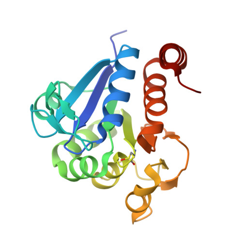
9YCU - PubMed Abstract:
Hydrogen bonds (H-bonds) are central to biomolecular structure and dynamics. Although H-bonds are typically characterized by well-defined proton positions, proton delocalization can play a key role in facilitating enzyme catalysis and allostery in some systems. Experimentally locating protons is difficult, hampering the study of the proton mobility in H-bonds. We used neutron crystallography and large quantum mechanics/molecular mechanics-Born Oppenheimer molecular dynamics (QM/MM-BOMD) simulations of human DJ-1 and its bacterial homolog YajL to validate atomic resolution X-ray crystallographic bond length analysis, directly visualizing the shared deuteron in a low-barrier hydrogen bond (LBHB) between Glu14-Asp23 in YajL that is a conventional H-bond in DJ-1. In addition, X-ray bond length analysis of protiated and perdeuterated DJ-1 and YajL shows no significant effect of deuteron substitution on these carboxylic acid-carboxylate H-bonds but does reveal an effect at the active site glutamic acid. Residues in the vicinity of Glu14-Asp23 that might favor LBHB formation in YajL were interrogated by mutagenesis of homologous residues in DJ-1. X-ray bond length analysis and QM/MM-BOMD simulations demonstrate that a distal I21T DJ-1 substitution increases proton delocalization in the Glu-Asp H-bond. In addition, simulations show that the extent of proton mobility in the H-bond influences correlated dimer-spanning motions in YajL and DJ-1. In total, we show that mutations within extended H-bonding networks can modulate proton transfer barriers in carboxylic acid-carboxylate H-bonds, allowing proton delocalization to be engineered using combined bioinformatic, structural, and computational information.
- Department of Biochemistry, University of Nebraska, Lincoln, NE, USA.
Organizational Affiliation: