9YBI | pdb_00009ybi

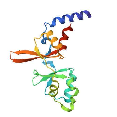
Structure of the CBS pair domain from zebrafish magnesium transporter CNNM2


Experimental Data Snapshot

  • Method: X-RAY DIFFRACTION
  • Resolution: 3.00 Å
  • R-Value Free: 
    0.253 (Depositor), 0.253 (DCC) 
  • R-Value Work: 
    0.235 (Depositor), 0.237 (DCC) 
  • R-Value Observed: 
    0.236 (Depositor) 

Starting Model: in silico
View more details

wwPDB Validation 3D Report Full Report

Validation slider image for 9YBI

This is version 1.1 of the entry. See complete history. 

Literature

The CNNM-binding interface drives PRL-3-dependent tumor progression in de novo cancer models.

Jolly, J.T., Felisbino, K., Mahbub, L., Al-Hamaly, M.A., Jin, M., Smith, C.N., Kozlov, G., Gehring, K., Blackburn, J.S.

(2026) iScience 29: 117338-117338

  • DOI: https://doi.org/10.1016/j.isci.2026.117338
  • Primary Citation Related Structures: 
    9YBI

  • PubMed Abstract: 

    Phosphatase of regenerating liver 3 (PRL-3) is a cancer-associated protein linked to tumor progression, metastasis, and poor patient outcomes, but the mechanism underlying its oncogenic activity remains unresolved. PRL-3 functions both as a phosphatase and as a binding partner of CNNM magnesium transporters, raising the question of which activity drives cancer progression. Here, we combine structure-guided separation-of-function mutants with biochemical and structural analyses, de novo zebrafish models of rhabdomyosarcoma and T cell acute lymphoblastic leukemia, and complementary human cancer cell assays to distinguish these mechanisms. PRL-3 wild type and the phosphatase-deficient, CNNM-binding-competent C104D mutant enhanced magnesium retention, stress tolerance, tumor-associated cell behaviors, tumor growth, and leukemia progression, whereas the CNNM-binding-deficient R138E mutant did not. These findings identify the CNNM-binding interface as a central mechanistic requirement for PRL-3-driven malignancy and suggest that therapeutic strategies focused solely on catalytic inhibition may not fully suppress its oncogenic functions.


  • Organizational Affiliation: 
    • Department of Molecular and Cellular Biochemistry, College of Medicine, University of Kentucky, Lexington, KY, USA.

Macromolecule Content 

  • Total Structure Weight: 35.93 kDa 
  • Atom Count: 2,251 
  • Modeled Residue Count: 296 
  • Deposited Residue Count: 316 
  • Unique protein chains: 1

Macromolecules

Find similar proteins by:|  3D Structure
Entity ID: 1
MoleculeChains  Sequence LengthOrganismDetailsImage
Metal transporter
A, B
158Danio rerioMutation(s): 0 
Gene Names: cnnm2a, im:7136247, si:ch211-67n3.4
UniProt
Find proteins for A2ATX7 (Danio rerio)
Explore A2ATX7 
Go to UniProtKB:  A2ATX7
Entity Groups
Sequence Clusters30% Identity50% Identity70% Identity90% Identity95% Identity100% Identity
UniProt GroupA2ATX7
Sequence Annotations
Expand
Reference Sequence

Experimental Data & Validation

Experimental Data

  • Method: X-RAY DIFFRACTION
  • Resolution: 3.00 Å
  • R-Value Free:  0.253 (Depositor), 0.253 (DCC) 
  • R-Value Work:  0.235 (Depositor), 0.237 (DCC) 
  • R-Value Observed: 0.236 (Depositor) 
Space Group: I 21 21 21
Unit Cell:
Length ( Å )Angle ( ˚ )
a = 86.907α = 90
b = 147.942β = 90
c = 151.897γ = 90
Software Package:
Software NamePurpose
PHENIXrefinement
HKL-2000data reduction
HKL-2000data scaling
PHENIXphasing

Structure Validation

View Full Validation Report


Entry History 

& Funding Information

Deposition Data


Funding OrganizationLocationGrant Number
Canadian Institutes of Health Research (CIHR)Canada--

Revision History  (Full details and data files)

  • Version 1.0: 2025-09-24
    Type: Initial release
  • Version 1.1: 2026-10-07
    Changes: Database references