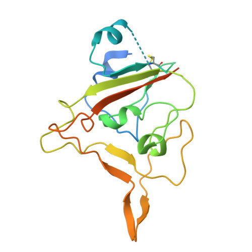
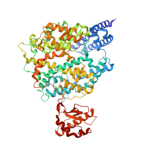
9W77 | pdb_00009w77

Rhinolophus cornutus Bat ACE2 Dimer in Complex with Two Rc-o319 Sarbecovirus Spike RBDs.


Experimental Data Snapshot

  • Method: ELECTRON MICROSCOPY
  • Resolution: 2.40 Å
  • Aggregation State: PARTICLE 
  • Reconstruction Method: SINGLE PARTICLE 

wwPDB Validation 3D Report Full Report

Validation slider image for 9W77

This is version 1.1 of the entry. See complete history. 

Literature

Structural basis for sarbecovirus Rc-o319 spike adaptation to Rhinolophus cornutus Bat ACE2 and constraints on switching to human ACE2.

Wang, J., Li, Z., Ma, Y., Li, Z., Yuan, H., Niu, C., Teng, Y., Liu, B., Li, M., Zhou, M., Liu, W., Feng, H., Chen, J., He, J., Chen, X., Xiong, X.

(2026) PLoS Pathog 22: e1014245-e1014245

  • DOI: https://doi.org/10.1371/journal.ppat.1014245
  • Primary Citation Related Structures: 
    9W75, 9W76, 9W77, 9W78

  • PubMed Abstract: 

    Bat sarbecoviruses often exhibit species-dependent ACE2 specificity. Understanding the determinants of receptor specificity enables better assessment of the cross-species transmission potential of sarbecoviruses. Here, we characterize the S-protein of Rc-o319, a sarbecovirus identified in Japanese Rhinolophus cornutus bats. Featuring an unusual 9-amino-acid deletion within its receptor binding motif (RBM), Rc-o319 S-protein utilizes its cognate R. cornutus ACE2 (bACE2R.cor) but not human ACE2 (hACE2), demonstrating highly restricted receptor specificity. Cryo-EM structures reveal two locked prefusion conformations of the Rc-o319 S-trimer and define a novel type of receptor-binding domain (RBD), featuring a distinct beta-loop (BL) within the RBM due to the RBM-deletion. The Rc-o319-RBD:bACE2R.cor complex structure reveals unique interactions mediated by the specialized BL and RBM-loop of Rc-o319-RBD and by a bACE2R.cor glycan. Structure-guided mutagenesis demonstrates that changes in BL and RBM-loop within the Rc-o319 S-RBD must occur simultaneously to allow medium-to-high-affinity hACE2 binding. Comparative assays further show that the bACE2R.cor receptor supports only a subset of sarbecoviruses, highlighting its restricted sarbecovirus compatibility. Our findings establish the Rc-o319 S-protein as a structurally and functionally specialized adaptation to R. cornutus ACE2 and identify the structural constraints limiting its cross-species transmission potential.


  • Organizational Affiliation: 
    • State Key Laboratory of Respiratory Disease, Guangdong Provincial Key Laboratory of Stem Cell and Regenerative Medicine, Guangdong-Hong Kong Joint Laboratory for Stem Cell and Regenerative Medicine, Guangzhou Institutes of Biomedicine and Health, Chinese Academy of Sciences, Guangzhou, China.

Macromolecule Content 

  • Total Structure Weight: 223.74 kDa 
  • Atom Count: 14,270 
  • Modeled Residue Count: 1,695 
  • Deposited Residue Count: 1,890 
  • Unique protein chains: 2

Macromolecules

Find similar proteins by:|  3D Structure
Entity ID: 1
MoleculeChains  Sequence LengthOrganismDetailsImage
Spike protein S1A [auth B],
D
214SarbecovirusMutation(s): 0 
Entity Groups
Sequence Clusters30% Identity50% Identity70% Identity90% Identity95% Identity100% Identity
Sequence Annotations
Expand
Reference Sequence
Find similar proteins by:|  3D Structure
Entity ID: 2
MoleculeChains  Sequence LengthOrganismDetailsImage
Angiotensin-converting enzymeB [auth C],
C [auth A]
731Rhinolophus cornutusMutation(s): 0 
Gene Names: RcACE2
EC: 3.4
UniProt
Find proteins for A0A7R7AIC6 (Rhinolophus cornutus)
Explore A0A7R7AIC6 
Go to UniProtKB:  A0A7R7AIC6
Entity Groups
Sequence Clusters30% Identity50% Identity70% Identity90% Identity95% Identity100% Identity
UniProt GroupA0A7R7AIC6
Glycosylation
Glycosylation Sites: 6
Sequence Annotations
Expand
Reference Sequence

Oligosaccharides

Help  
Entity ID: 3
MoleculeChains Length2D Diagram GlycosylationD Interactions
2-acetamido-2-deoxy-beta-D-glucopyranose-(1-4)-2-acetamido-2-deoxy-beta-D-glucopyranose
E, F, G, I, J
E, F, G, I, J, K
2N-Glycosylation
Glycosylation Resources
GlyTouCan: G42666HT
GlyCosmos: G42666HT
GlyGen: G42666HT
Entity ID: 4
MoleculeChains Length2D Diagram GlycosylationD Interactions
alpha-D-mannopyranose-(1-2)-alpha-D-mannopyranose-(1-3)-beta-D-mannopyranose-(1-4)-2-acetamido-2-deoxy-beta-D-glucopyranose-(1-4)-2-acetamido-2-deoxy-beta-D-glucopyranose
H
5N-Glycosylation
Glycosylation Resources
GlyTouCan: G42227JK
GlyCosmos: G42227JK
GlyGen: G42227JK
Entity ID: 5
MoleculeChains Length2D Diagram GlycosylationD Interactions
alpha-D-mannopyranose-(1-3)-[alpha-D-mannopyranose-(1-6)]beta-D-mannopyranose-(1-4)-2-acetamido-2-deoxy-beta-D-glucopyranose-(1-4)-2-acetamido-2-deoxy-beta-D-glucopyranose
L
5N-Glycosylation
Glycosylation Resources
GlyTouCan: G22768VO
GlyCosmos: G22768VO
GlyGen: G22768VO

Experimental Data & Validation

Experimental Data

  • Method: ELECTRON MICROSCOPY
  • Resolution: 2.40 Å
  • Aggregation State: PARTICLE 
  • Reconstruction Method: SINGLE PARTICLE 
EM Software:
TaskSoftware PackageVersion
MODEL REFINEMENTPHENIX
RECONSTRUCTIONcryoSPARC

Structure Validation

View Full Validation Report


Entry History 

& Funding Information

Deposition Data


Funding OrganizationLocationGrant Number
National Natural Science Foundation of China (NSFC)China82341085

Revision History  (Full details and data files)

  • Version 1.0: 2025-10-29
    Type: Initial release
  • Version 1.1: 2026-11-18
    Changes: Data collection, Database references