9VN5 | pdb_00009vn5

Structure of WDR5 in complex with MBD3C R43K


Experimental Data Snapshot

  • Method: X-RAY DIFFRACTION
  • Resolution: 1.90 Å
  • R-Value Free: 
    0.231 (Depositor), 0.233 (DCC) 
  • R-Value Work: 
    0.205 (Depositor), 0.209 (DCC) 
  • R-Value Observed: 
    0.206 (Depositor) 

Starting Model: experimental
View more details

wwPDB Validation   3D Report Full Report


This is version 1.1 of the entry. See complete history


Literature

Structural insights into lysine-mediated WIN site recognition reveal an alternative "AK" motif for WDR5 binding.

Pan, Y.Li, H.Chen, L.Sun, X.Xu, L.Yang, Y.

(2025) Biochem Biophys Res Commun 779: 152440-152440

  • DOI: https://doi.org/10.1016/j.bbrc.2025.152440
  • Primary Citation of Related Structures:  
    9VN5

  • PubMed Abstract: 

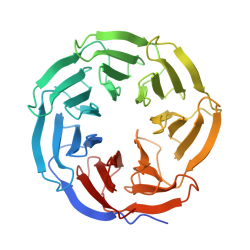
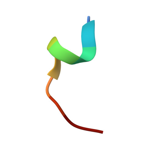
    WD repeat-containing protein 5 (WDR5) is a highly conserved chromatin-associated scaffold protein that recognizes short, arginine-containing WDR5 interacting (WIN) motifs in various partners to assemble transcriptional complexes. Although the WIN motif has been considered strictly arginine-dependent, our previous binding assays showed that the MBD3C_R43K variant retained binding to WDR5 (K D  = 1.31 μM), though the underlying mechanism remained unclear. Here, we report the crystal structure of WDR5 in complex with MBD3C 40-51 (R43K) peptide at 1.9 Å resolution, revealing that lysine can also insert into the WIN pocket. Structural analysis shows that the lysine side chain mimics the canonical arginine interactions, forming hydrogen bonds with Ser91and engaging in hydrophobic contacts with Ser49, Phe133, Cys261, and Ile305 within the conserved WIN-binding site of WDR5. Interestingly, this experimentally determined structure contrasts with AlphaFold3 predictions, which incorrectly placed Arg45 into the WIN pocket rather than Lys43, underscoring the limitations of current predictive models for protein-peptide complexes involving mutations. Overall, our findings redefine the WIN motif consensus to include an alternative "AK" motif and highlight the contributions of residues at positions +2, +3, and +4 in stabilizing WDR5 binding. This work broadens the understanding of WDR5 substrate recognition and offers a structural framework for designing WIN-site inhibitors that target lysine-mediated interactions.


  • Organizational Affiliation
    • School of Life Sciences, Anhui University, Hefei, Anhui, 230601, China.

Macromolecules
Find similar proteins by:  (by identity cutoff)  |  3D Structure
Entity ID: 1
MoleculeChains Sequence LengthOrganismDetailsImage
WD repeat-containing protein 5A,
C [auth B]
315Homo sapiensMutation(s): 0 
Gene Names: WDR5BIG3
UniProt & NIH Common Fund Data Resources
Find proteins for P61964 (Homo sapiens)
Explore P61964 
Go to UniProtKB:  P61964
PHAROS:  P61964
GTEx:  ENSG00000196363 
Entity Groups  
Sequence Clusters30% Identity50% Identity70% Identity90% Identity95% Identity100% Identity
UniProt GroupP61964
Sequence Annotations
Expand
  • Reference Sequence

Find similar proteins by:  Sequence   |   3D Structure  

Entity ID: 2
MoleculeChains Sequence LengthOrganismDetailsImage
Methyl-CpG binding domain protein 3B [auth C],
D
10Mus musculusMutation(s): 1 
Gene Names: Mbd3
UniProt
Find proteins for D3YTR4 (Mus musculus)
Explore D3YTR4 
Go to UniProtKB:  D3YTR4
Entity Groups  
Sequence Clusters30% Identity50% Identity70% Identity90% Identity95% Identity100% Identity
UniProt GroupD3YTR4
Sequence Annotations
Expand
  • Reference Sequence
Experimental Data & Validation

Experimental Data

  • Method: X-RAY DIFFRACTION
  • Resolution: 1.90 Å
  • R-Value Free:  0.231 (Depositor), 0.233 (DCC) 
  • R-Value Work:  0.205 (Depositor), 0.209 (DCC) 
  • R-Value Observed: 0.206 (Depositor) 
Space Group: C 1 2 1
Unit Cell:
Length ( Å )Angle ( ˚ )
a = 116.265α = 90
b = 47.376β = 113.34
c = 129.472γ = 90
Software Package:
Software NamePurpose
PHENIXrefinement
PDB_EXTRACTdata extraction
autoPXdata reduction
Aimlessdata scaling
PHASERphasing

Structure Validation

View Full Validation Report


Entry History & Funding Information

Deposition Data


Funding OrganizationLocationGrant Number
Not funded--

Revision History  (Full details and data files)

  • Version 1.0: 2025-08-13
    Type: Initial release
  • Version 1.1: 2025-08-20
    Changes: Database references