9VMA | pdb_00009vma

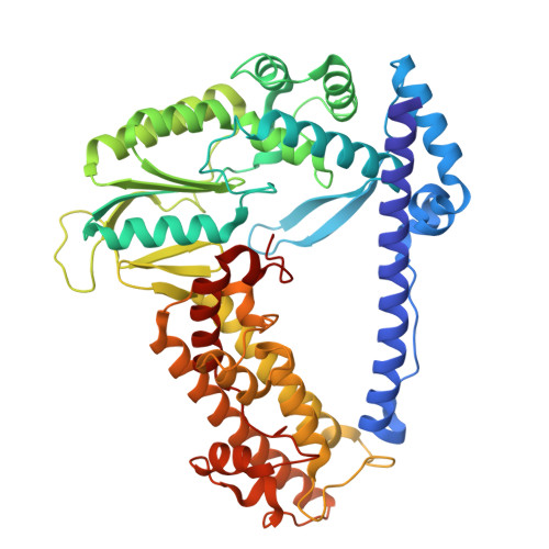
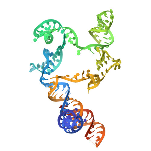
Cryo-EM structure of substrate-bound DRT9 hexamer complex


Experimental Data Snapshot

  • Method: ELECTRON MICROSCOPY
  • Resolution: 3.46 Å
  • Aggregation State: PARTICLE 
  • Reconstruction Method: SINGLE PARTICLE 

wwPDB Validation 3D Report Full Report

Validation slider image for 9VMA

This is version 1.2 of the entry. See complete history. 

Literature

Non-coding RNA mediates the defense-associated reverse transcriptase (DRT) anti-phage oligomerization transition.

Han, J., Liu, B., Tang, J., Zhang, S., Wang, X., Li, X., Zhang, Q., Liu, Z., Wang, W., Liu, Y., Zhou, R., Yin, H., Wei, Y., Li, Z., Zhang, M., Deng, Z., Zhang, H.

(2025) EMBO J 44: 5429-5442

  • DOI: https://doi.org/10.1038/s44318-025-00544-8
  • Primary Citation Related Structures: 
    9VKU, 9VMA

  • PubMed Abstract: 

    Defense-associated reverse transcriptase (DRT) systems are implicated in prokaryotic resistance to viral infections, yet the molecular mechanisms underlying their functionality remain largely unknown. Here, we characterize a two-component DRT9 system, composed of a reverse transcriptase (RT) and a non-coding RNA (ncRNA), which exhibits a protein-primed DNA synthesis activity upon phage infection. We also determine its cryo-electron microscopy (cryo-EM) structures in different functional states. DRT9 RT binds to ncRNA, forming a dimer of dimers configuration that assembles into a trimer of dimers upon substrate binding. This oligomerization transition, crucial for DRT9-mediated anti-phage defense, is facilitated by a ncRNA cooperative self-assembly manner. Furthermore, substrate binding induces large conformational movements around the catalytic pocket of DRT9 RT, revealing a "lock-switch" mechanism for enzymatic activation. Notably, phylogenetic analysis and functional assays identify a unique N-terminal helix extension required for ncRNA stabilization and enzymatic activity, distinct from previously reported reverse transcriptase systems. Overall, our findings illuminate the molecular basis of DRT9-mediated antiviral defense and expand the functional and mechanistic diversity of the DRT family.


  • Organizational Affiliation: 
    • Key Laboratory of Immune Microenvironment and Disease (Ministry of Education), State Key Laboratory of Experimental Hematology, Tianjin Medical University Cancer Institute and Hospital, The Province and Ministry Co-sponsored Collaborative Innovation Center for Medical Epigenetics, Tianjin Institute of Immunology, School of Basic Medical Sciences, Tianjin Medical University, Tianjin, China.

Macromolecule Content 

  • Total Structure Weight: 722.57 kDa 
  • Atom Count: 45,606 
  • Modeled Residue Count: 3,963 
  • Deposited Residue Count: 4,146 
  • Unique protein chains: 1
  • Unique nucleic acid chains: 2

Macromolecules


Find similar proteins by:|  3D Structure
Entity ID: 1
MoleculeChains  Sequence LengthOrganismDetailsImage
RNA-dependent DNA polymerase
A, B, C, D, E
A, B, C, D, E, F
499Escherichia coliMutation(s): 0 
Gene Names: GQA06_05700
UniProt
Find proteins for A0A6D0I497 (Escherichia coli)
Explore A0A6D0I497 
Go to UniProtKB:  A0A6D0I497
Entity Groups
Sequence Clusters30% Identity50% Identity70% Identity90% Identity95% Identity100% Identity
UniProt GroupA0A6D0I497
Sequence Annotations
Expand
Reference Sequence
Find similar nucleic acids by:  (by identity cutoff) 
Entity ID: 2
MoleculeChains LengthOrganismImage
RNA (188-MER)
G, H, I, J, K
G, H, I, J, K, L
188Escherichia coli
Sequence Annotations
Expand
Reference Sequence
Find similar nucleic acids by:  Sequence
Entity ID: 3
MoleculeChains LengthOrganismImage
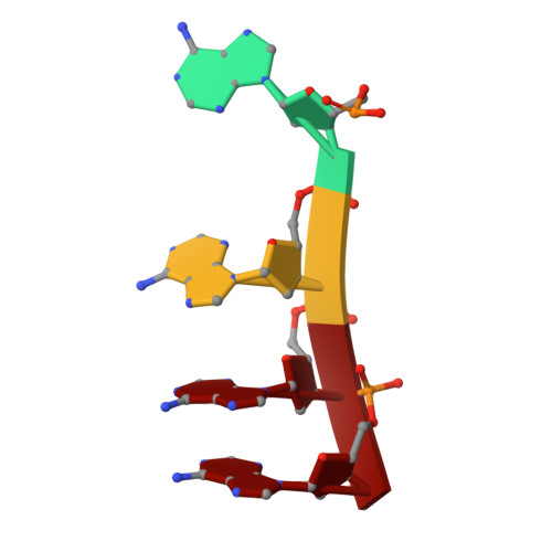
DNA (5'-D(P*AP*AP*AP*A)-3')
M, N, O, P, Q
M, N, O, P, Q, R
4Escherichia coli
Sequence Annotations
Expand
Reference Sequence

Small Molecules

Ligands 2 Unique
IDChains Name / Formula / InChI Key2D Diagram3D Interactions
DTP

Query on DTP


Download:Ideal Coordinates CCD File
AA [auth E]
CA [auth F]
S [auth A]
U [auth B]
W [auth C]
AA [auth E],
CA [auth F],
S [auth A],
U [auth B],
W [auth C],
Y [auth D]
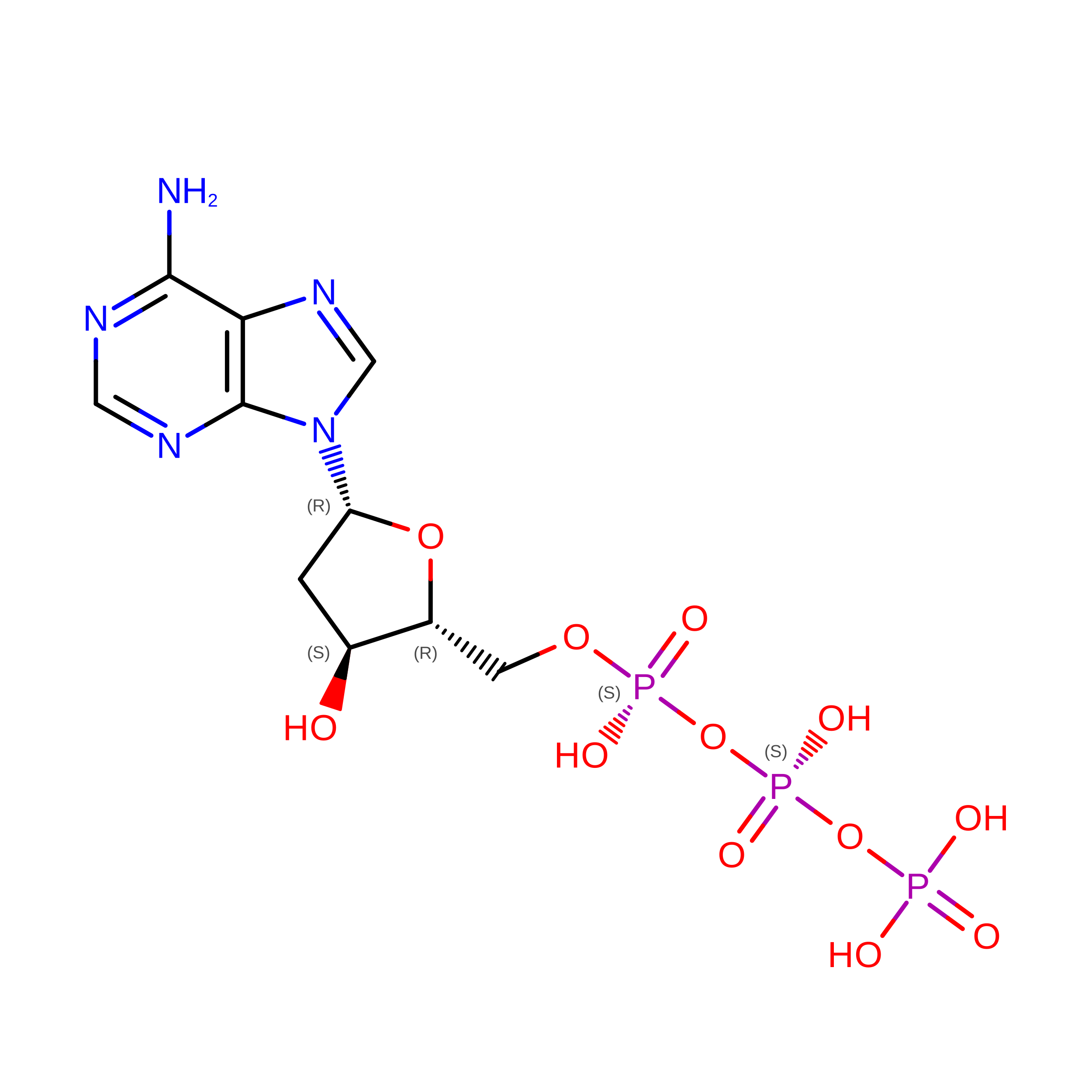
2'-DEOXYADENOSINE 5'-TRIPHOSPHATE
C10 H16 N5 O12 P3
SUYVUBYJARFZHO-RRKCRQDMSA-N
MG

Query on MG


Download:Ideal Coordinates CCD File
BA [auth E]
DA [auth F]
EA [auth G]
FA [auth H]
GA [auth I]
BA [auth E],
DA [auth F],
EA [auth G],
FA [auth H],
GA [auth I],
HA [auth J],
IA [auth K],
JA [auth L],
T [auth A],
V [auth B],
X [auth C],
Z [auth D]
MAGNESIUM ION
Mg
JLVVSXFLKOJNIY-UHFFFAOYSA-N

Experimental Data & Validation

Experimental Data

  • Method: ELECTRON MICROSCOPY
  • Resolution: 3.46 Å
  • Aggregation State: PARTICLE 
  • Reconstruction Method: SINGLE PARTICLE 
EM Software:
TaskSoftware PackageVersion
MODEL REFINEMENTPHENIX1.20.1_4487
RECONSTRUCTIONcryoSPARC

Structure Validation

View Full Validation Report


Entry History 

& Funding Information

Deposition Data


Funding OrganizationLocationGrant Number
Other government--

Revision History  (Full details and data files)

  • Version 1.0: 2025-08-20
    Type: Initial release
  • Version 1.1: 2025-09-03
    Changes: Data collection, Database references, Structure summary
  • Version 1.2: 2025-10-15
    Changes: Data collection, Database references