9VH1 | pdb_00009vh1

cryoEM structure of ptuA-ptuB complex in Retron-Eco7 anti-phage system


Experimental Data Snapshot

  • Method: ELECTRON MICROSCOPY
  • Resolution: 3.40 Å
  • Aggregation State: PARTICLE 
  • Reconstruction Method: SINGLE PARTICLE 

wwPDB Validation   3D Report Full Report


This is version 1.0 of the entry. See complete history


Literature

Phage nuclease-mediated defense activation of the bacterial Retron-Eco7 toxin-antitoxin system.

Dai, Z.Liu, C.Wang, Y.Chen, X.Fu, X.Yang, K.Zhu, R.Jia, X.Chen, Y.Tao, P.Guan, Z.Zou, T.

(2025) Nucleic Acids Res 53

  • DOI: https://doi.org/10.1093/nar/gkaf1173
  • Primary Citation of Related Structures:  
    9VH1, 9VHE

  • PubMed Abstract: 

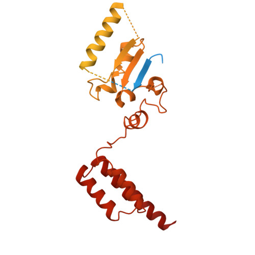
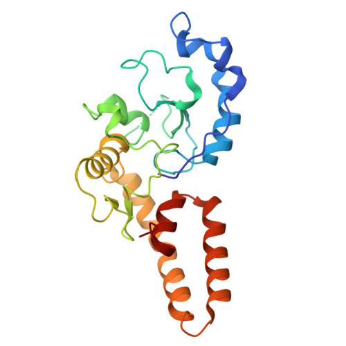
    Retrons are bacterial antiphage defense systems comprising a reverse transcriptase (RT), a non-coding RNA (ncRNA), and cognate effector proteins. The RT synthesizes multicopy single-stranded DNA (msDNA) from the ncRNA template to detect phage invasion. This study focuses on Retron-Eco7, which integrates retron-based sensing with the effector module of Septu-a characterized antiphage system in which the PtuAB complex mediates nuclease-dependent defense. However, the activation mechanism of this hybrid system remains unclear. Here, we determined cryo-electron microscopy structures of the RT-msDNA-PtuAB quaternary complex and the PtuAB binary complex in Retron-Eco7. Structural analyses reveal that the DNA stem-loop of msDNA extensively interacts with PtuA subunits via electrostatic interactions. We establish Retron-Eco7 as a novel toxin-antitoxin system, in which RT-msDNA acts as the antitoxin, directly binding and neutralizing the PtuAB toxin. Furthermore, we identified a phage-encoded flap endonuclease as a trigger for Retron-Eco7 activation, which cleaves msDNA to release the PtuAB toxin. Our findings demonstrate the diversity in bacterial retron defense systems and uncover a novel activation mechanism of the Septu-derived retron toxin-antitoxin system.


  • Organizational Affiliation
    • National Key Laboratory of Agricultural Microbiology, Hubei Hongshan Laboratory, Huazhong Agricultural University, Wuhan 430070, China.

Macromolecules
Find similar proteins by:  (by identity cutoff)  |  3D Structure
Entity ID: 1
MoleculeChains Sequence LengthOrganismDetailsImage
Retron Ec78 probable ATPaseA [auth D],
B [auth E]
550Escherichia coliMutation(s): 0 
Gene Names: Ga0100609_101823
UniProt
Find proteins for P0DV91 (Escherichia coli)
Explore P0DV91 
Go to UniProtKB:  P0DV91
Entity Groups  
Sequence Clusters30% Identity50% Identity70% Identity90% Identity95% Identity100% Identity
UniProt GroupP0DV91
Sequence Annotations
Expand
  • Reference Sequence
Find similar proteins by:  (by identity cutoff)  |  3D Structure
Entity ID: 2
MoleculeChains Sequence LengthOrganismDetailsImage
Retron Ec78 putative HNH endonucleaseC [auth F]216Escherichia coliMutation(s): 0 
Gene Names: Ga0100609_101824
UniProt
Find proteins for P0DV92 (Escherichia coli)
Explore P0DV92 
Go to UniProtKB:  P0DV92
Entity Groups  
Sequence Clusters30% Identity50% Identity70% Identity90% Identity95% Identity100% Identity
UniProt GroupP0DV92
Sequence Annotations
Expand
  • Reference Sequence
Small Molecules
Experimental Data & Validation

Experimental Data

  • Method: ELECTRON MICROSCOPY
  • Resolution: 3.40 Å
  • Aggregation State: PARTICLE 
  • Reconstruction Method: SINGLE PARTICLE 
EM Software:
TaskSoftware PackageVersion
MODEL REFINEMENTPHENIX1.21.2_5419
RECONSTRUCTIONcryoSPARC

Structure Validation

View Full Validation Report


Entry History & Funding Information

Deposition Data


Funding OrganizationLocationGrant Number
National Natural Science Foundation of China (NSFC)China--

Revision History  (Full details and data files)

  • Version 1.0: 2025-12-03
    Type: Initial release