9V7J | pdb_00009v7j

Phycobilisome core from Gloeobacter violaceus PCC 7421


Experimental Data Snapshot

  • Method: ELECTRON MICROSCOPY
  • Resolution: 2.85 Å
  • Aggregation State: PARTICLE 
  • Reconstruction Method: SINGLE PARTICLE 

wwPDB Validation   3D Report Full Report


This is version 1.1 of the entry. See complete history


Literature

Structure and quenching of a bundle-shaped phycobilisome.

Burtseva, A.D.Slonimskiy, Y.B.Baymukhametov, T.N.Sinetova, M.A.Gvozdev, D.A.Tsoraev, G.V.Cherepanov, D.A.Maksimov, E.G.Popov, V.O.Boyko, K.M.Sluchanko, N.N.

(2025) Sci Adv 11: eadz6774-eadz6774

  • DOI: https://doi.org/10.1126/sciadv.adz6774
  • Primary Citation of Related Structures:  
    9V7G, 9V7H, 9V7I, 9V7J, 9V7K, 9V7L

  • PubMed Abstract: 

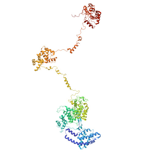
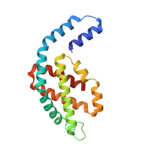
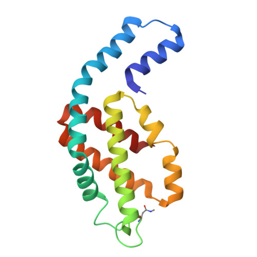
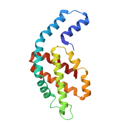
    Cyanobacteria use soluble antenna megacomplexes, phycobilisomes (PBSs), to maximize light-harvesting efficiency and small photoswitchable orange carotenoid proteins (OCPs) to down-regulate PBSs in high light. Among known PBS morphologies, the one from the basal cyanobacterial genus Gloeobacter still lacks detailed structural characterization. Here, we reconstructed a cryo-electron microscopy structure of the >10-megadalton Gloeobacter violaceus PBS, with diverging, conformationally mobile bundles of rods composed of stacked phycoerythrin and phycocyanin hexamers, stemming from a pentacylindrical allophycocyanin core belted by auxiliary phycocyanin hexamers. We show how two Gloeobacter -specific multidomain linker proteins, Glr1262 and Glr2806, maintain this bundle-shaped architecture and reveal its differential regulation via nonphotochemical quenching by two OCP types of G. violaceus that recognize separate binding sites within the allophycocyanin core, including lateral cylinders absent in tricylindrical cores.


  • Organizational Affiliation
    • A.N. Bach Institute of Biochemistry, Federal Research Center of Biotechnology of the Russian Academy of Sciences, Moscow 119071, Russia.

Macromolecules
Find similar proteins by:  (by identity cutoff)  |  3D Structure
Entity ID: 1
MoleculeChains Sequence LengthOrganismDetailsImage
Phycobiliprotein ApcEA,
B [auth C]
1,155Gloeobacter violaceus PCC 7421Mutation(s): 0 
UniProt
Find proteins for Q7NL81 (Gloeobacter violaceus (strain ATCC 29082 / PCC 7421))
Explore Q7NL81 
Go to UniProtKB:  Q7NL81
Entity Groups  
Sequence Clusters30% Identity50% Identity70% Identity90% Identity95% Identity100% Identity
UniProt GroupQ7NL81
Sequence Annotations
Expand
  • Reference Sequence
Find similar proteins by:  (by identity cutoff)  |  3D Structure
Entity ID: 2
MoleculeChains Sequence LengthOrganismDetailsImage
Allophycocyanin alpha subunit161Gloeobacter violaceus PCC 7421Mutation(s): 0 
UniProt
Find proteins for Q7NL80 (Gloeobacter violaceus (strain ATCC 29082 / PCC 7421))
Explore Q7NL80 
Go to UniProtKB:  Q7NL80
Entity Groups  
Sequence Clusters30% Identity50% Identity70% Identity90% Identity95% Identity100% Identity
UniProt GroupQ7NL80
Sequence Annotations
Expand
  • Reference Sequence
Find similar proteins by:  (by identity cutoff)  |  3D Structure
Entity ID: 3
MoleculeChains Sequence LengthOrganismDetailsImage
Allophycocyanin beta subunit161Gloeobacter violaceus PCC 7421Mutation(s): 0 
UniProt
Find proteins for Q7NL79 (Gloeobacter violaceus (strain ATCC 29082 / PCC 7421))
Explore Q7NL79 
Go to UniProtKB:  Q7NL79
Entity Groups  
Sequence Clusters30% Identity50% Identity70% Identity90% Identity95% Identity100% Identity
UniProt GroupQ7NL79
Sequence Annotations
Expand
  • Reference Sequence
Find similar proteins by:  (by identity cutoff)  |  3D Structure
Entity ID: 4
MoleculeChains Sequence LengthOrganismDetailsImage
Phycobilisome core componentQA [auth s],
SA [auth u]
161Gloeobacter violaceus PCC 7421Mutation(s): 0 
UniProt
Find proteins for Q7NJA2 (Gloeobacter violaceus (strain ATCC 29082 / PCC 7421))
Explore Q7NJA2 
Go to UniProtKB:  Q7NJA2
Entity Groups  
Sequence Clusters30% Identity50% Identity70% Identity90% Identity95% Identity100% Identity
UniProt GroupQ7NJA2
Sequence Annotations
Expand
  • Reference Sequence
Find similar proteins by:  (by identity cutoff)  |  3D Structure
Entity ID: 5
MoleculeChains Sequence LengthOrganismDetailsImage
Phycobilisome 7.8 kDa linker polypeptide, allophycocyanin-associated, core69Gloeobacter violaceus PCC 7421Mutation(s): 0 
UniProt
Find proteins for Q7NL78 (Gloeobacter violaceus (strain ATCC 29082 / PCC 7421))
Explore Q7NL78 
Go to UniProtKB:  Q7NL78
Entity Groups  
Sequence Clusters30% Identity50% Identity70% Identity90% Identity95% Identity100% Identity
UniProt GroupQ7NL78
Sequence Annotations
Expand
  • Reference Sequence
Small Molecules
Ligands 1 Unique
IDChains Name / Formula / InChI Key2D Diagram3D Interactions
CYC (Subject of Investigation/LOI)
Query on CYC

Download Ideal Coordinates CCD File 
AD [auth Q]
AE [auth q]
AF [auth AG]
BD [auth R]
BE [auth r]
AD [auth Q],
AE [auth q],
AF [auth AG],
BD [auth R],
BE [auth r],
BF [auth AH],
CD [auth S],
CE [auth s],
CF [auth AI],
DD [auth T],
DE [auth t],
DF [auth AJ],
ED [auth U],
EE [auth u],
EF [auth AK],
FD [auth V],
FE [auth v],
FF [auth AL],
GD [auth W],
GE [auth w],
GF [auth AM],
HD [auth X],
HE [auth x],
HF [auth AN],
ID [auth Y],
IE [auth y],
IF [auth AO],
JD [auth Z],
JE [auth z],
JF [auth AP],
KD [auth a],
KE [auth 1],
KF [auth AQ],
LD [auth b],
LE [auth 2],
LF [auth AR],
MC [auth A],
MD [auth c],
ME [auth 3],
MF [auth AS],
NC [auth C],
ND [auth d],
NE [auth 4],
NF [auth AT],
OC [auth M],
OD [auth e],
OE [auth 5],
OF [auth AU],
PC [auth N],
PD [auth f],
PE [auth 6],
PF [auth AV],
QC [auth O],
QD [auth g],
QE [auth 7],
QF [auth AW],
RC [auth P],
RD [auth h],
RE [auth 8],
RF [auth AX],
SC [auth E],
SD [auth i],
SE [auth 9],
TC [auth F],
TD [auth j],
TE [auth 0],
UC [auth G],
UD [auth k],
UE [auth AA],
VC [auth H],
VD [auth l],
VE [auth AB],
WC [auth I],
WD [auth m],
WE [auth AC],
XC [auth J],
XD [auth n],
XE [auth AD],
YC [auth K],
YD [auth o],
YE [auth AE],
ZC [auth L],
ZD [auth p],
ZE [auth AF]
PHYCOCYANOBILIN
C33 H40 N4 O6
VXTXPYZGDQPMHK-GMXXPEQVSA-N
Modified Residues  1 Unique
IDChains TypeFormula2D DiagramParent
MEN
Query on MEN
BA [auth d]
BB [auth 4]
BC [auth AT]
D [auth N]
DA [auth f]
BA [auth d],
BB [auth 4],
BC [auth AT],
D [auth N],
DA [auth f],
DB [auth 6],
DC [auth AV],
F [auth P],
FA [auth h],
FB [auth 8],
FC [auth AX],
H [auth F],
HA [auth j],
HB [auth 0],
J [auth H],
JA [auth l],
JB [auth AB],
L [auth J],
LA [auth n],
LB [auth AD],
N [auth L],
NA [auth p],
NB [auth AF],
OA [auth q],
P [auth R],
PB [auth AH],
R [auth T],
RB [auth AJ],
T [auth V],
TA [auth v],
TB [auth AL],
V [auth X],
VA [auth x],
VB [auth AN],
X [auth Z],
XA [auth z],
XB [auth AP],
Z [auth b],
ZA [auth 2],
ZB [auth AR]
L-PEPTIDE LINKINGC5 H10 N2 O3ASN
Experimental Data & Validation

Experimental Data

  • Method: ELECTRON MICROSCOPY
  • Resolution: 2.85 Å
  • Aggregation State: PARTICLE 
  • Reconstruction Method: SINGLE PARTICLE 
EM Software:
TaskSoftware PackageVersion
MODEL REFINEMENTREFMAC5.8.0425

Structure Validation

View Full Validation Report


Entry History & Funding Information

Deposition Data


Funding OrganizationLocationGrant Number
Russian Science FoundationRussian Federation23-74-00062

Revision History  (Full details and data files)

  • Version 1.0: 2025-09-24
    Type: Initial release
  • Version 1.1: 2025-10-29
    Changes: Data collection, Database references