9UVI | pdb_00009uvi

Crystal structure of beta 2 microglobulin in complex with antibody BBM.1


Experimental Data Snapshot

  • Method: X-RAY DIFFRACTION
  • Resolution: 2.50 Å
  • R-Value Free: 
    0.246 (Depositor), 0.245 (DCC) 
  • R-Value Work: 
    0.197 (Depositor), 0.197 (DCC) 
  • R-Value Observed: 
    0.202 (Depositor) 

Starting Model: experimental
View more details

wwPDB Validation   3D Report Full Report


This is version 1.1 of the entry. See complete history


Literature

Crystal structure of the beta 2 -microglobulin-BBM.1 antibody complex reveals the molecular basis of antigen recognition.

Wu, J.Zeng, F.Wang, X.Wei, P.

(2025) Acta Crystallogr D Struct Biol 81: 473-481

  • DOI: https://doi.org/10.1107/S2059798325006370
  • Primary Citation of Related Structures:  
    9UVI

  • PubMed Abstract: 

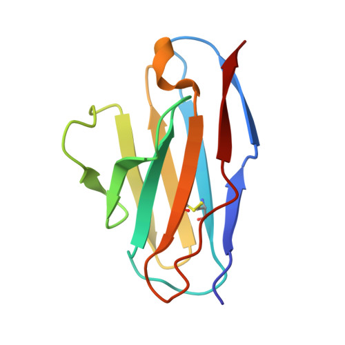
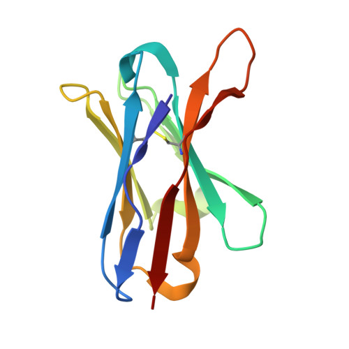
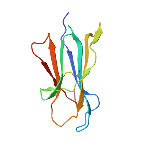
    β 2 -Microglobulin (β 2 M) is an essential component of major histocompatibility complex class I (MHC-I) molecules, with a well established canonical role in immune surveillance. Beyond its classical functions, accumulating evidence has highlighted β 2 M as a multifaceted biomarker, with elevated serum levels closely associated with disease burden and prognosis in metabolic disorders, malignancies, autoimmune diseases and central nervous system conditions. In this study, we resolved the crystal structure of human β 2 M in complex with the mouse monoclonal antibody BBM.1 at 2.50 Å resolution using X-ray crystallography. Structural analysis revealed that BBM.1 binds β 2 M through multiple CDRs, recognizing key surface residues including Glu36, Asp38, Lys41, Asn42, Glu44, Arg45, Glu47 and Arg81. The interaction is anchored by a central hydrophobic core formed by Trp32 (light chain), Trp99 (heavy chain) and Ile92 (β 2 M), which is deeply buried in the interface. Surrounding this core is a well organized polar interaction network composed of hydrogen bonds and salt bridges, primarily involving β 2 M residues Lys41, Glu44, Arg45 and Glu47. Notably, the Arg45 residue deeply embeds into the antibody-binding pocket, forming several crucial interactions. These findings not only validate previous biochemical and mutational data but also identify new epitope residues, providing a structural foundation for the development and optimization of precision therapeutic strategies targeting β 2 M.


  • Organizational Affiliation
    • Guangxi Key Laboratory of Special Biomedicine, School of Medicine, Guangxi University, Nanning 530004, People's Republic of China.

Macromolecules
Find similar proteins by:  (by identity cutoff)  |  3D Structure
Entity ID: 1
MoleculeChains Sequence LengthOrganismDetailsImage
BBM.1 light chain107Mus musculusMutation(s): 0 
Entity Groups  
Sequence Clusters30% Identity50% Identity70% Identity90% Identity95% Identity100% Identity
Sequence Annotations
Expand
  • Reference Sequence
Find similar proteins by:  (by identity cutoff)  |  3D Structure
Entity ID: 2
MoleculeChains Sequence LengthOrganismDetailsImage
BBM.1 heavy chain116Mus musculusMutation(s): 0 
Entity Groups  
Sequence Clusters30% Identity50% Identity70% Identity90% Identity95% Identity100% Identity
Sequence Annotations
Expand
  • Reference Sequence
Find similar proteins by:  (by identity cutoff)  |  3D Structure
Entity ID: 3
MoleculeChains Sequence LengthOrganismDetailsImage
Beta-2-microglobulin99Homo sapiensMutation(s): 0 
Gene Names: B2MCDABP0092HDCMA22P
UniProt & NIH Common Fund Data Resources
Find proteins for P61769 (Homo sapiens)
Explore P61769 
Go to UniProtKB:  P61769
PHAROS:  P61769
GTEx:  ENSG00000166710 
Entity Groups  
Sequence Clusters30% Identity50% Identity70% Identity90% Identity95% Identity100% Identity
UniProt GroupP61769
Sequence Annotations
Expand
  • Reference Sequence
Experimental Data & Validation

Experimental Data

  • Method: X-RAY DIFFRACTION
  • Resolution: 2.50 Å
  • R-Value Free:  0.246 (Depositor), 0.245 (DCC) 
  • R-Value Work:  0.197 (Depositor), 0.197 (DCC) 
  • R-Value Observed: 0.202 (Depositor) 
Space Group: P 41 21 2
Unit Cell:
Length ( Å )Angle ( ˚ )
a = 59.99α = 90
b = 59.99β = 90
c = 187.81γ = 90
Software Package:
Software NamePurpose
PHENIXrefinement
MOSFLMdata reduction
Aimlessdata scaling
PHASERphasing

Structure Validation

View Full Validation Report


Entry History & Funding Information

Deposition Data


Funding OrganizationLocationGrant Number
National Natural Science Foundation of China (NSFC)China--

Revision History  (Full details and data files)

  • Version 1.0: 2025-09-03
    Type: Initial release
  • Version 1.1: 2025-09-17
    Changes: Database references