9US5 | pdb_00009us5

Domain N deletion mutant of Klebsiella pneumoniae maltohexaose-producing alpha-amylase in complex with maltohexaose


Experimental Data Snapshot

  • Method: X-RAY DIFFRACTION
  • Resolution: 2.66 Å
  • R-Value Free: 
    0.226 (Depositor), 0.227 (DCC) 
  • R-Value Work: 
    0.182 (Depositor), 0.185 (DCC) 

Starting Model: experimental
View more details

wwPDB Validation 3D Report Full Report

Validation slider image for 9US5

This is version 1.1 of the entry. See complete history

Literature

Crystal structure of Klebsiella pneumoniae maltohexaose-producing alpha-amylase.

Fujimoto, Z.Kishine, N.Momma, M.

(2025) J Biochem 178: 201-207

  • DOI: https://doi.org/10.1093/jb/mvaf034
  • Primary Citation Related Structures: 
    9US3, 9US4, 9US5, 9US6

  • PubMed Abstract: 

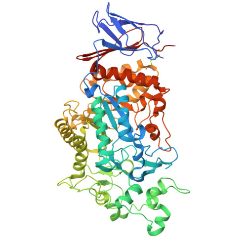
    The α-amylase from Klebsiella pneumoniae (KpAmy13), which belongs to glycoside hydrolase family 13 subfamily 19, produces maltohexaose as an initial product when acting on starch and has been characterized as a maltohexaose-producing α-amylase. The crystal structure of KpAmy13 was determined at a resolution of 1.9 Å, revealing the structures of all its domains: N, A, B, and C. Domain N resembles the starch-binding domain known as carbohydrate-binding module family 69, found in α-glucan-related proteins. Although domain N does not conserve the starch-binding residues observed in other proteins, it has several hydrophobic residues on its surface, which might be involved in promoting catalysis. The catalytic cleft is located at the bottom of a circular depression. The domain N-truncated mutant of KpAmy13 in complex with maltohexaose showed that its non-reducing end glucose docks at subsite -6. The long and complex structure of domain B contributes to forming a cleft of the right size for six glucose moieties, demonstrating the exo-acting mechanism.


  • Organizational Affiliation
    • Research Center for Advanced Analysis, National Agriculture and Food Research Organization, 2-1-2 Kannondai, Tsukuba, Ibaraki 305-8518, Japan.

Macromolecule Content 

  • Total Structure Weight: 64.54 kDa 
  • Atom Count: 4,560 
  • Modeled Residue Count: 558 
  • Deposited Residue Count: 566 
  • Unique protein chains: 1

Macromolecules

Find similar proteins by:|  3D Structure
Entity ID: 1
MoleculeChains  Sequence LengthOrganismDetailsImage
Maltohexaose-producing amylase566Klebsiella pneumoniaeMutation(s): 0 
EC: 3.2.1.98
UniProt
Find proteins for Q9RHR1 (Klebsiella pneumoniae)
Explore Q9RHR1 
Go to UniProtKB:  Q9RHR1
Entity Groups
Sequence Clusters30% Identity50% Identity70% Identity90% Identity95% Identity100% Identity
UniProt GroupQ9RHR1
Sequence Annotations
Expand
Reference Sequence

Oligosaccharides

Help  
Entity ID: 2
MoleculeChains Length2D Diagram GlycosylationD Interactions
alpha-D-glucopyranose-(1-4)-alpha-D-glucopyranose-(1-4)-alpha-D-glucopyranose-(1-4)-alpha-D-glucopyranose
B
4N/A
Glycosylation Resources
GlyTouCan: G87171PZ
GlyCosmos: G87171PZ
GlyGen: G87171PZ

Biologically Interesting Molecules (External Reference) 

1 Unique

Experimental Data & Validation

Experimental Data

  • Method: X-RAY DIFFRACTION
  • Resolution: 2.66 Å
  • R-Value Free:  0.226 (Depositor), 0.227 (DCC) 
  • R-Value Work:  0.182 (Depositor), 0.185 (DCC) 
Space Group: I 4 2 2
Unit Cell:
Length ( Å )Angle ( ˚ )
a = 163.084α = 90
b = 163.084β = 90
c = 139.376γ = 90
Software Package:
Software NamePurpose
REFMACrefinement
XDSdata reduction
SCALAdata scaling
MOLREPphasing

Structure Validation

View Full Validation Report


Entry History 

& Funding Information

Deposition Data


Funding OrganizationLocationGrant Number
Not funded--

Revision History  (Full details and data files)

  • Version 1.0: 2025-07-09
    Type: Initial release
  • Version 1.1: 2025-09-03
    Changes: Database references