9UKV | pdb_00009ukv

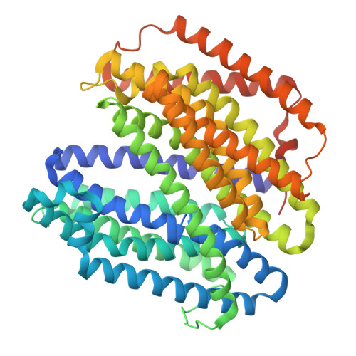
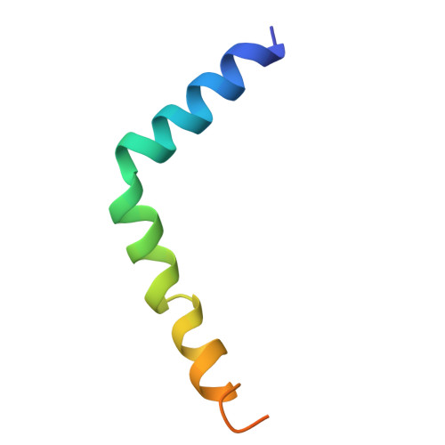
JM Complex - E. coli MurJ, Levivirus M lysis protein LysM (SglM)


Experimental Data Snapshot

  • Method: ELECTRON MICROSCOPY
  • Resolution: 3.05 Å
  • Aggregation State: PARTICLE 
  • Reconstruction Method: SINGLE PARTICLE 

wwPDB Validation 3D Report Full Report

Validation slider image for 9UKV

This is version 1.1 of the entry. See complete history. 

Literature

Phage lysis protein Lys M acts as a wedge to block MurJ conformational changes.

Kohga, H., Lertpreedakorn, N., Miyazaki, R., Wu, S., Hosoda, K., Tanaka, H., Takahashi, Y.S., Yoshikaie, K., Kuruma, Y., Shigematsu, H., Mori, T., Tsukazaki, T.

(2025) Sci Adv 11: eady8083-eady8083

  • DOI: https://doi.org/10.1126/sciadv.ady8083
  • Primary Citation Related Structures: 
    9UKV

  • PubMed Abstract: 

    Many antibiotics target essential cellular processes. To combat multidrug-resistant bacteria, new antibacterial strategies are needed. In the peptidoglycan biogenesis pathway in Escherichia coli , MurJ, the lipid II flippase, is an essential membrane protein. The 37-residue protein M from the Levivirus phage, known as Lys M or Sgl M , targets MurJ and induces cell lysis; however, its molecular mechanism remains unclear. Here, we present the cryo-EM structure of the MurJ/Lys M (JM) complex at 3.09-angstrom resolution, revealing that Lys M interacts with the crevasse between TM2 and TM7 of MurJ, locking MurJ in an outward-facing conformation, with Lys M acting like a wedge. Alanine-scanning mutagenesis and pull-down assays revealed key residues responsible for Lys M function, and molecular dynamics simulations showed that Lys M stabilizes MurJ's outward-facing state. These findings demonstrate an unprecedented phage-derived mechanism for blocking lipid II transport, providing a structural framework for designing MurJ-targeted antimicrobial agents.


  • Organizational Affiliation: 
    • Nara Institute of Science and Technology, Ikoma, Nara, Japan.

Macromolecule Content 

  • Total Structure Weight: 63.64 kDa 
  • Atom Count: 4,188 
  • Modeled Residue Count: 548 
  • Deposited Residue Count: 581 
  • Unique protein chains: 2

Macromolecules

Find similar proteins by:|  3D Structure
Entity ID: 1
MoleculeChains  Sequence LengthOrganismDetailsImage
Lipid II flippase MurJ534Escherichia coliMutation(s): 0 
Gene Names: murJ, mviN, yceN, b1069, JW1056
Membrane Entity: Yes 
UniProt
Find proteins for P0AF16 (Escherichia coli (strain K12))
Explore P0AF16 
Go to UniProtKB:  P0AF16
Entity Groups
Sequence Clusters30% Identity50% Identity70% Identity90% Identity95% Identity100% Identity
UniProt GroupP0AF16
Sequence Annotations
Expand
Reference Sequence
Find similar proteins by:|  3D Structure
Entity ID: 2
MoleculeChains  Sequence LengthOrganismDetailsImage
Lysis protein47Enterobacteria phage MMutation(s): 0 
Gene Names: lys
UniProt
Find proteins for K7QK87 (Enterobacteria phage M)
Explore K7QK87 
Go to UniProtKB:  K7QK87
Entity Groups
Sequence Clusters30% Identity50% Identity70% Identity90% Identity95% Identity100% Identity
UniProt GroupK7QK87
Sequence Annotations
Expand
Reference Sequence

Experimental Data & Validation

Experimental Data

  • Method: ELECTRON MICROSCOPY
  • Resolution: 3.05 Å
  • Aggregation State: PARTICLE 
  • Reconstruction Method: SINGLE PARTICLE 
EM Software:
TaskSoftware PackageVersion
MODEL REFINEMENTPHENIX

Structure Validation

View Full Validation Report


Entry History 

& Funding Information

Deposition Data


Funding OrganizationLocationGrant Number
Japan Society for the Promotion of Science (JSPS)Japan--

Revision History  (Full details and data files)

  • Version 1.0: 2025-09-10
    Type: Initial release
  • Version 1.1: 2026-03-04
    Changes: Data collection, Database references