9SI1 | pdb_00009si1

Mouse otoferlin (216-1931) in the lipid-free Ca2+-bound state, "closed-like" conformation


Experimental Data Snapshot

  • Method: ELECTRON MICROSCOPY
  • Resolution: 3.10 Å
  • Aggregation State: PARTICLE 
  • Reconstruction Method: SINGLE PARTICLE 

Starting Model: experimental
View more details

wwPDB Validation 3D Report Full Report

Validation slider image for 9SI1

This is version 1.0 of the entry. See complete history

Literature

Structure and function of otoferlin, a synaptic protein of sensory hair cells essential for hearing.

Chen, H.Cretu, C.Trebilcock, A.Evdokimova, N.Babai, N.Feldmann, L.Leidner, F.Benseler, F.Mutschall, S.Esch, K.Szabo, C.Z.K.Pena, V.Pape, C.Grubmuller, H.Strenzke, N.Brose, N.Wichmann, C.Preobraschenski, J.Moser, T.

(2025) Sci Adv 11: eady8532-eady8532

  • DOI: https://doi.org/10.1126/sciadv.ady8532
  • Primary Citation Related Structures: 
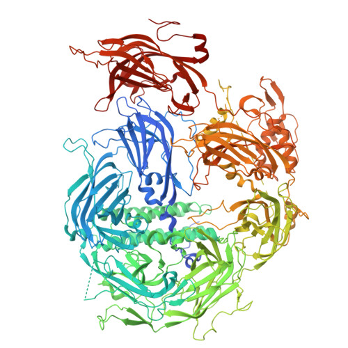
    9QE2, 9SE5, 9SEA, 9SEG, 9SFL, 9SH0, 9SI1

  • PubMed Abstract: 

    Hearing relies upon speedy synaptic transmission of sound information from inner hair cells (IHCs) to spiral ganglion neurons. To accomplish this, IHCs use a sophisticated presynaptic machinery including the multi-C 2 domain protein otoferlin that is affected by human deafness mutations. Otoferlin is essential for IHC exocytosis, but how it binds Ca 2+ and the target membrane to serve synaptic vesicle (SV) tethering, docking, and fusion remained unclear. Here, we obtained cryo-electron microscopy structures of otoferlin and employed molecular dynamics simulations of membrane binding. We show that membrane binding by otoferlin involves C 2 B-C 2 G domains and repositions C 2 F and C 2 G domains. Disruption of Ca 2+ -binding sites of the C 2 D domain in mice altered synaptic sound encoding and eliminated the Ca 2+ cooperativity of IHC exocytosis, indicating that it requires the binding of several Ca 2+ -ions by otoferlin. Together, our findings elucidate molecular mechanisms underlying otoferlin-mediated SV docking and support the role of otoferlin as Ca 2+ sensor of SV fusion in IHCs.


  • Organizational Affiliation
    • Institute for Auditory Neuroscience and InnerEarLab, University Medical Center Göttingen, Göttingen, Germany.

Macromolecule Content 

  • Total Structure Weight: 200.78 kDa 
  • Atom Count: 12,135 
  • Modeled Residue Count: 1,510 
  • Deposited Residue Count: 1,767 
  • Unique protein chains: 1

Macromolecules

Find similar proteins by:|  3D Structure
Entity ID: 1
MoleculeChains  Sequence LengthOrganismDetailsImage
Otoferlin1,767Mus musculusMutation(s): 0 
Gene Names: Otof
UniProt
Find proteins for A0A0R4J1K2 (Mus musculus)
Explore A0A0R4J1K2 
Go to UniProtKB:  A0A0R4J1K2
Entity Groups
Sequence Clusters30% Identity50% Identity70% Identity90% Identity95% Identity100% Identity
UniProt GroupA0A0R4J1K2
Sequence Annotations
Expand
Reference Sequence

Small Molecules

Ligands 1 Unique
IDChains Name / Formula / InChI Key2D Diagram3D Interactions
CA
(Subject of Investigation/LOI)

Query on CA


Download:Ideal Coordinates CCD File
B [auth A]
C [auth A]
D [auth A]
E [auth A]
F [auth A]
B [auth A],
C [auth A],
D [auth A],
E [auth A],
F [auth A],
G [auth A],
H [auth A],
I [auth A],
J [auth A]
CALCIUM ION
Ca
BHPQYMZQTOCNFJ-UHFFFAOYSA-N

Experimental Data & Validation

Experimental Data

  • Method: ELECTRON MICROSCOPY
  • Resolution: 3.10 Å
  • Aggregation State: PARTICLE 
  • Reconstruction Method: SINGLE PARTICLE 
EM Software:
TaskSoftware PackageVersion
RECONSTRUCTIONcryoSPARCv.4.7
MODEL REFINEMENTPHENIX1.21.2_5419

Structure Validation

View Full Validation Report


Entry History 

& Funding Information

Deposition Data


Funding OrganizationLocationGrant Number
German Research Foundation (DFG)GermanyCR 937/2-1

Revision History  (Full details and data files)

  • Version 1.0: 2025-11-05
    Type: Initial release