A Luminescence-Based Screening Platform for Lanthanide-Binding Peptides and Proteins.
Klassen, R., Heider, A., Kugler, H., Groll, M., Zeymer, C.(2025) ACS Chem Biol 20: 2897-2906
- PubMed: 41248128 Search on PubMed
- DOI: https://doi.org/10.1021/acschembio.5c00670
- Primary Citation Related Structures:
9S7R - PubMed Abstract:
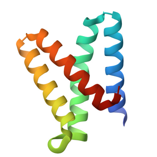
The specific incorporation of lanthanide ions is a promising strategy to equip biomolecules with a new function. Their long-lived luminescence, strong anomalous X-ray scattering, paramagnetism, Lewis acidity, and photoredox activity are attractive features for protein-based probes, materials, and catalysts. However, natural lanthanide-binding proteins are rare, and de novo design is often complicated by unspecific binding to negatively charged patches on protein surfaces. We thus aimed to develop an efficient workflow to screen libraries of protein scaffolds for their ability to coordinate lanthanides. Here, we introduce a microtiter plate-based assay, which employs commercial filter plates and a dual readout based on sensitized Tb 3+ luminescence. We first benchmarked our procedure using control proteins with and without lanthanide-binding sites, demonstrating that site-specific coordination and surface binding can be distinguished. The stringency of this protocol also allowed screening for small lanthanide-binding peptides in the presence of a large expression tag. We then designed a de novo scaffold library derived from a helical bundle protein and applied our screening platform. We could identify lanthanide-binding variants with nanomolar affinity, distinct lanthanide specificity, and increased thermostability in response to metal binding. Our approach will support the discovery and evolution of lanthanide-binding peptides and proteins for various applications in vitro and in living cells.
- Center for Functional Protein Assemblies & Department of Bioscience, TUM School of Natural Sciences, Technical University of Munich (TUM), Garching 85748, Germany.
Organizational Affiliation: