9RMS | pdb_00009rms

GluA4 in complex with TARP-2, Resting II state, structure of TMD/LBD domains


Experimental Data Snapshot

  • Method: ELECTRON MICROSCOPY
  • Resolution: 3.80 Å
  • Aggregation State: PARTICLE 
  • Reconstruction Method: SINGLE PARTICLE 

Starting Models: experimental
View more details

wwPDB Validation   3D Report Full Report


This is version 1.2 of the entry. See complete history


Literature

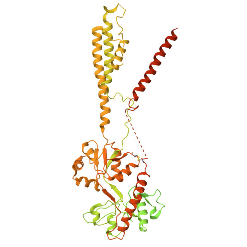
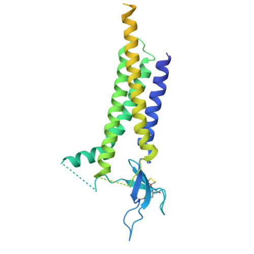
GluA4 AMPA receptor gating mechanisms and modulation by auxiliary proteins.

Vega-Gutierrez, C.Picanol-Parraga, J.Sanchez-Valls, I.Ribon-Fuster, V.D.P.Soto, D.Herguedas, B.

(2025) Nat Struct Mol Biol 32: 2416-2428

  • DOI: https://doi.org/10.1038/s41594-025-01666-7
  • Primary Citation of Related Structures:  
    9IGZ, 9QDN, 9QPW, 9RMS, 9RMW, 9RN4, 9RN7, 9RNH

  • PubMed Abstract: 

    AMPA-type glutamate receptors, fundamental ion channels for fast excitatory neurotransmission and synaptic plasticity, contain a GluA tetrameric core surrounded by auxiliary proteins such as transmembrane AMPA receptor regulatory proteins (TARPs) or Cornichons. Their exact composition and stoichiometry govern functional properties, including kinetics, calcium permeability and trafficking. The GluA1-GluA3 subunits predominate in the adult forebrain and are well characterized. However, we lack structural information on full-length GluA4-containing AMPARs, a subtype that has specific roles in brain development and specific cell types in mammals. Here we present the cryo-electron microscopy structures of rat GluA4:TARP-γ2 trapped in active, resting and desensitized states, covering a full gating cycle. Additionally, we describe the structure of GluA4 alone, which displays a classical Y-shaped conformation. In resting conditions, GluA4:TARP-γ2 adopts two conformations, one resembling the desensitized states of other GluA subunits. Moreover, we identify a regulatory site for TARP-γ2 in the ligand-binding domain that modulates gating kinetics. Our findings uncover distinct features of GluA4, highlighting how subunit composition and auxiliary proteins shape receptor structure and dynamics, expanding glutamatergic signaling diversity.


  • Organizational Affiliation
    • Institute for Biocomputation and Physics of Complex Systems (BIFI), Department of Biochemistry and Molecular and Cell Biology, University of Zaragoza, Zaragoza, Spain.

Macromolecules
Find similar proteins by:  (by identity cutoff)  |  3D Structure
Entity ID: 1
MoleculeChains Sequence LengthOrganismDetailsImage
Isoform 2 of Glutamate receptor 4
A, B, C, D
882Rattus norvegicusMutation(s): 1 
Gene Names: Gria4Glur4
UniProt
Find proteins for P19493 (Rattus norvegicus)
Explore P19493 
Go to UniProtKB:  P19493
Entity Groups  
Sequence Clusters30% Identity50% Identity70% Identity90% Identity95% Identity100% Identity
UniProt GroupP19493
Sequence Annotations
Expand
  • Reference Sequence
Find similar proteins by:  (by identity cutoff)  |  3D Structure
Entity ID: 2
MoleculeChains Sequence LengthOrganismDetailsImage
Voltage-dependent calcium channel gamma-2 subunit
E, F, G, H
323Rattus norvegicusMutation(s): 1 
Gene Names: Cacng2Stg
UniProt
Find proteins for Q71RJ2 (Rattus norvegicus)
Explore Q71RJ2 
Go to UniProtKB:  Q71RJ2
Entity Groups  
Sequence Clusters30% Identity50% Identity70% Identity90% Identity95% Identity100% Identity
UniProt GroupQ71RJ2
Sequence Annotations
Expand
  • Reference Sequence
Experimental Data & Validation

Experimental Data

  • Method: ELECTRON MICROSCOPY
  • Resolution: 3.80 Å
  • Aggregation State: PARTICLE 
  • Reconstruction Method: SINGLE PARTICLE 
EM Software:
TaskSoftware PackageVersion
MODEL REFINEMENTPHENIX1.19.2_4158:
RECONSTRUCTIONcryoSPARC4.3.1

Structure Validation

View Full Validation Report


Entry History & Funding Information

Deposition Data


Funding OrganizationLocationGrant Number
Ministerio de Ciencia e Innovacion (MCIN)SpainPID2019-106284GA-I00

Revision History  (Full details and data files)

  • Version 1.0: 2025-09-24
    Type: Initial release
  • Version 1.1: 2025-10-01
    Changes: Data collection, Database references
  • Version 1.2: 2025-12-24
    Changes: Data collection, Database references