


SARS-CoV-2 3CL protease with a bound inhibitor
Mac Sweeney, A.To be published.
Experimental Data Snapshot
Starting Model: experimental
View more details
Entity ID: 1 | |||||
|---|---|---|---|---|---|
| Molecule | Chains | Sequence Length | Organism | Details | Image |
| 3C-like proteinase nsp5 | 306 | Severe acute respiratory syndrome coronavirus 2 | Mutation(s): 0 EC: 3.4.22.69 | 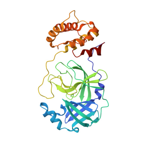 | |
UniProt | |||||
Entity Groups | |||||
| Sequence Clusters | 30% Identity50% Identity70% Identity90% Identity95% Identity100% Identity | ||||
| UniProt Group | P0DTC1 | ||||
Sequence AnnotationsExpand | |||||
Reference Sequence | |||||
| Ligands 1 Unique | |||||
|---|---|---|---|---|---|
| ID | Chains | Name / Formula / InChI Key | 2D Diagram | 3D Interactions | |
| A1JF7 (Subject of Investigation/LOI) Download:Ideal Coordinates CCD File | C [auth A], D [auth B] | ~{N}-(4-bromanyl-3-nitro-phenyl)-2-(1,2,3-triazol-1-yl)ethanamide C10 H8 Br N5 O3 JWYPHVGKUYHWNL-UHFFFAOYSA-N |  | ||
| Length ( Å ) | Angle ( ˚ ) |
|---|---|
| a = 67.91 | α = 90 |
| b = 100.24 | β = 90 |
| c = 104.01 | γ = 90 |
| Software Name | Purpose |
|---|---|
| REFMAC | refinement |
| autoPROC | data reduction |
| autoPROC | data scaling |
| PHASER | phasing |
| Funding Organization | Location | Grant Number |
|---|---|---|
| Not funded | -- |


