9R4Z | pdb_00009r4z

Murine AA amyloid fibril morphology III (AA III)


Experimental Data Snapshot

  • Method: ELECTRON MICROSCOPY
  • Resolution: 3.50 Å
  • Aggregation State: HELICAL ARRAY 
  • Reconstruction Method: HELICAL 

wwPDB Validation   3D Report Full Report


This is version 1.1 of the entry. See complete history


Literature

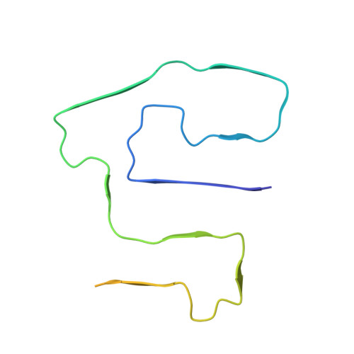
Cryo-EM Observation of AA Amyloid Fibrils in Mouse Model of Systemic AApoAII Amyloidosis.

Andreotti, G.Higuchi, K.Schmidt, M.Fandrich, M.

(2025) J Mol Biology 437: 169438-169438

  • DOI: https://doi.org/10.1016/j.jmb.2025.169438
  • Primary Citation of Related Structures:  
    9R4Z

  • PubMed Abstract: 

    The co-deposition of amyloid fibrils from different precursor proteins is a topic of increasing relevance for protein misfolding diseases. Using cryo-electron microscopy (cryo-EM), we here determined the structures of two serum amyloid A (SAA) protein-derived amyloid fibril morphologies that were extracted from a mouse strain that is primarily known to be associated with apolipoprotein A-II-derived amyloid fibrils. The two fibril morphologies show the same protomer conformation as in previously reported ex vivo amyloid fibrils from SAA protein but a different relative arrangement of fibril protein stacks. These data establish that serum amyloid A-derived amyloid fibrils share the same fibril protein fold in different mouse strains and disease contexts.


  • Organizational Affiliation
    • Institute of Protein Biochemistry, Ulm University, 89081 Ulm, Germany. Electronic address: giada.andreotti@uni-ulm.de.

Macromolecules
Find similar proteins by:  (by identity cutoff)  |  3D Structure
Entity ID: 1
MoleculeChains Sequence LengthOrganismDetailsImage
Serum amyloid A-2 protein103Mus musculusMutation(s): 0 
UniProt
Find proteins for P05367 (Mus musculus)
Explore P05367 
Go to UniProtKB:  P05367
Entity Groups  
Sequence Clusters30% Identity50% Identity70% Identity90% Identity95% Identity100% Identity
UniProt GroupP05367
Sequence Annotations
Expand
  • Reference Sequence
Experimental Data & Validation

Experimental Data

  • Method: ELECTRON MICROSCOPY
  • Resolution: 3.50 Å
  • Aggregation State: HELICAL ARRAY 
  • Reconstruction Method: HELICAL 
EM Software:
TaskSoftware PackageVersion
RECONSTRUCTIONRELION5.0
MODEL REFINEMENTCoot

Structure Validation

View Full Validation Report


Entry History & Funding Information

Deposition Data


Funding OrganizationLocationGrant Number
German Research Foundation (DFG)Germany1279/Z03
German Research Foundation (DFG)GermanyA03

Revision History  (Full details and data files)

  • Version 1.0: 2025-10-01
    Type: Initial release
  • Version 1.1: 2025-10-08
    Changes: Data collection, Database references