9QWD | pdb_00009qwd

X-ray structure of furin (PCSK3) in complex with the biphenyl-derived compound 34 (mi2470)


Experimental Data Snapshot

  • Method: X-RAY DIFFRACTION
  • Resolution: 1.60 Å
  • R-Value Free: 
    0.161 (Depositor), 0.167 (DCC) 
  • R-Value Work: 
    0.135 (Depositor), 0.145 (DCC) 
  • R-Value Observed: 
    0.136 (Depositor) 

Starting Model: experimental
View more details

wwPDB Validation 3D Report Full Report

Validation slider image for 9QWD

Ligand Structure Quality Assessment 


This is version 1.0 of the entry. See complete history

Literature

Design, Synthesis, and Characterization of Dichlorobiphenyl-Derived Inhibitors of the Proprotein Convertase Furin.

Lange, R.W.Boller, C.Loresch, M.Bloch, K.Bottcher-Friebertshauser, E.Brandstetter, H.Dahms, S.O.Steinmetzer, T.

(2025) J Med Chem 68: 25157-25170

  • DOI: https://doi.org/10.1021/acs.jmedchem.5c02157
  • Primary Citation Related Structures: 
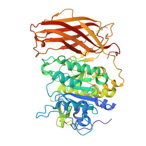
    9QWB, 9QWC, 9QWD, 9QWE, 9QWF, 9QWG

  • PubMed Abstract: 

    The proprotein convertase (PC) furin emerged as promising drug target for the treatment of numerous infectious diseases, cancer and cystic fibrosis. A recently described nonpeptidic lead structure served as template to develop a new series of PC inhibitors containing a dichlorobiphenyl-derived core segment decorated with a left and right inhibitor arm. The compounds were tested for their inhibitory potency against furin and the structurally related PC7. The most potent compounds inhibited furin with K i values <5 nM, whereas most of them were significantly weaker inhibitors of PC7. Only for one compound, a significant potency with a K i value of 7.3 nM against PC7 was found. Furthermore, crystal structures of six inhibitors in complex with furin were determined. Selected inhibitors were additionally tested for their antiviral potency against the furin-dependent H7N7 influenza A strain SC35M; a significant antiviral potency was found for compound 9 .


  • Organizational Affiliation
    • Institute of Pharmaceutical Chemistry, Philipps University, Marbacher Weg 10, D-35032 Marburg, Germany.

Macromolecule Content 

  • Total Structure Weight: 53.9 kDa 
  • Atom Count: 4,359 
  • Modeled Residue Count: 472 
  • Deposited Residue Count: 482 
  • Unique protein chains: 1

Macromolecules

Find similar proteins by:|  3D Structure
Entity ID: 1
MoleculeChains  Sequence LengthOrganismDetailsImage
Furin482Homo sapiensMutation(s): 0 
Gene Names: FURINFURPACEPCSK3
EC: 3.4.21.75
UniProt & NIH Common Fund Data Resources
Find proteins for P09958 (Homo sapiens)
Explore P09958 
Go to UniProtKB:  P09958
PHAROS:  P09958
GTEx:  ENSG00000140564 
Entity Groups
Sequence Clusters30% Identity50% Identity70% Identity90% Identity95% Identity100% Identity
UniProt GroupP09958
Glycosylation
Glycosylation Sites: 1Go to GlyGen: P09958-1
Sequence Annotations
Expand
Reference Sequence

Small Molecules

Ligands 6 Unique
IDChains Name / Formula / InChI Key2D Diagram3D Interactions
A1JA2
(Subject of Investigation/LOI)

Query on A1JA2


Download:Ideal Coordinates CCD File
Q [auth A]1-[[3-[3,5-bis(chloranyl)phenyl]-5-[(4-pyridin-4-ylpiperazin-1-yl)methyl]phenyl]methyl]-4-pyridin-4-yl-piperazine
C32 H34 Cl2 N6
IFPHOKXTHCEYJO-UHFFFAOYSA-N
NAG

Query on NAG


Download:Ideal Coordinates CCD File
P [auth A]2-acetamido-2-deoxy-beta-D-glucopyranose
C8 H15 N O6
OVRNDRQMDRJTHS-FMDGEEDCSA-N
DMS

Query on DMS


Download:Ideal Coordinates CCD File
J [auth A]
K [auth A]
L [auth A]
M [auth A]
N [auth A]
J [auth A],
K [auth A],
L [auth A],
M [auth A],
N [auth A],
O [auth A]
DIMETHYL SULFOXIDE
C2 H6 O S
IAZDPXIOMUYVGZ-UHFFFAOYSA-N
CA

Query on CA


Download:Ideal Coordinates CCD File
B [auth A],
C [auth A],
D [auth A]
CALCIUM ION
Ca
BHPQYMZQTOCNFJ-UHFFFAOYSA-N
CL

Query on CL


Download:Ideal Coordinates CCD File
I [auth A]CHLORIDE ION
Cl
VEXZGXHMUGYJMC-UHFFFAOYSA-M
NA

Query on NA


Download:Ideal Coordinates CCD File
E [auth A],
F [auth A],
G [auth A],
H [auth A]
SODIUM ION
Na
FKNQFGJONOIPTF-UHFFFAOYSA-N

Experimental Data & Validation

Experimental Data

  • Method: X-RAY DIFFRACTION
  • Resolution: 1.60 Å
  • R-Value Free:  0.161 (Depositor), 0.167 (DCC) 
  • R-Value Work:  0.135 (Depositor), 0.145 (DCC) 
  • R-Value Observed: 0.136 (Depositor) 
Space Group: P 65 2 2
Unit Cell:
Length ( Å )Angle ( ˚ )
a = 131.507α = 90
b = 131.507β = 90
c = 155.35γ = 120
Software Package:
Software NamePurpose
PHENIXrefinement
PHENIXphasing
XDSdata reduction
XDSdata scaling

Structure Validation

View Full Validation Report


Ligand Structure Quality Assessment 


Entry History 

& Funding Information

Deposition Data


Funding OrganizationLocationGrant Number
Austrian Science FundAustriaM 2730-B
Austrian Science FundAustriaP26_P36648

Revision History  (Full details and data files)

  • Version 1.0: 2025-12-24
    Type: Initial release