9QM9 | pdb_00009qm9

TMEM55B tandem RING-like domains in complex with RILPL1 C-terminal peptide


Experimental Data Snapshot

  • Method: X-RAY DIFFRACTION
  • Resolution: 1.55 Å
  • R-Value Free: 
    0.246 (Depositor), 0.246 (DCC) 
  • R-Value Work: 
    0.205 (Depositor), 0.205 (DCC) 
  • R-Value Observed: 
    0.207 (Depositor) 

Starting Model: experimental
View more details

wwPDB Validation 3D Report Full Report

Validation slider image for 9QM9

This is version 1.1 of the entry. See complete history

Literature

Structural basis for binding of RILPL1 to TMEM55B reveals a lysosomal platform for adaptor assembly through a conserved peptide motif.

Waschbusch, D.Pal, P.Nirujogi, R.S.Cavin, M.Singh, J.Alessi, D.R.Khan, A.R.

(2026) Structure 34: 296-310.e5

  • DOI: https://doi.org/10.1016/j.str.2025.11.003
  • Primary Citation Related Structures: 
    8OQH, 9QM9

  • PubMed Abstract: 

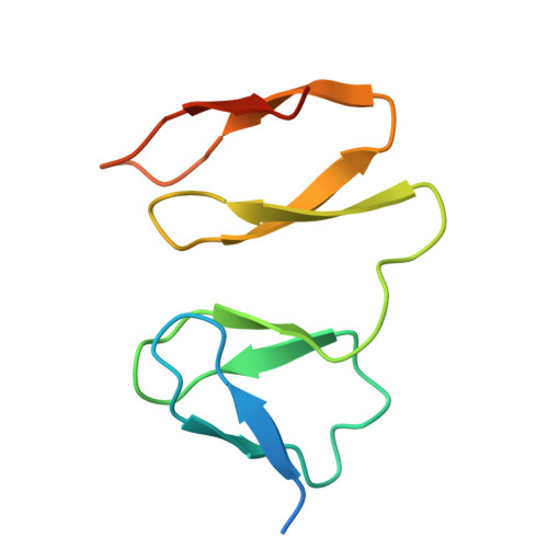
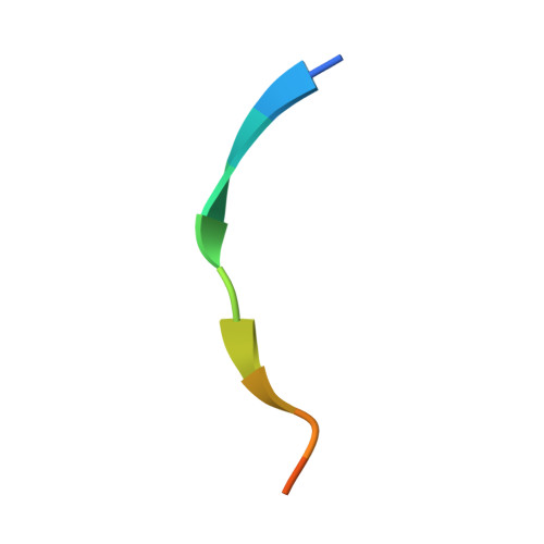
    Inherited mutations in VPS35 and LRRK2 kinase lead to hyperphosphorylation of Rab GTPases. RH2 domain-containing proteins from the RILP homology family, such as RILPL1, are Rab effectors that recognize the LRRK2-phosphorylated switch 2 threonine of phospho-Rab8A and phospho-Rab10. Phospho-Rabs are also seen on lysosomal membranes in complex with RILPL1 and TMEM55B, a 284-residue lysosomal membrane protein lacking homology to known proteins. Here, we report crystal structures of the cytosolic region 80-166 of TMEM55B alone and in complex with a C-terminal RILPL1 peptide, which we define as the TMEM55B-binding motif (TBM). The RILPL1 TBM sits in a shallow groove across two tandem RING-like domains of TMEM55B, each forming a Zn 2+ -stabilized 40-residue β-sandwich. Co-immunoprecipitation and mass spectrometry studies indicate that TMEM55B forms complexes independently of phospho-Rabs with conserved TBMs found in JIP3, JIP4, OCRL, WDR81, and TBC1D9B. These studies suggest that TMEM55B acts as a central hub for adaptor recruitment on lysosomes.


  • Organizational Affiliation
    • School of Biochemistry and Immunology, Trinity College Dublin, Dublin 2, Ireland.

Macromolecule Content 

  • Total Structure Weight: 21.36 kDa 
  • Atom Count: 1,403 
  • Modeled Residue Count: 167 
  • Deposited Residue Count: 195 
  • Unique protein chains: 2

Macromolecules

Find similar proteins by:|  3D Structure
Entity ID: 1
MoleculeChains  Sequence LengthOrganismDetailsImage
Type 1 phosphatidylinositol 4,5-bisphosphate 4-phosphatase
A, B
91Homo sapiensMutation(s): 2 
Gene Names: PIP4P1C14orf9TMEM55B
EC: 3.1.3.78
UniProt & NIH Common Fund Data Resources
Find proteins for Q86T03 (Homo sapiens)
Explore Q86T03 
Go to UniProtKB:  Q86T03
PHAROS:  Q86T03
GTEx:  ENSG00000165782 
Entity Groups
Sequence Clusters30% Identity50% Identity70% Identity90% Identity95% Identity100% Identity
UniProt GroupQ86T03
Sequence Annotations
Expand
Reference Sequence
Find similar proteins by:  Sequence   |   3D Structure  
Entity ID: 2
MoleculeChains  Sequence LengthOrganismDetailsImage
RILP-like protein 1C [auth D]13Homo sapiensMutation(s): 0 
Gene Names: RILPL1RLP1
UniProt & NIH Common Fund Data Resources
Find proteins for Q5EBL4 (Homo sapiens)
Explore Q5EBL4 
Go to UniProtKB:  Q5EBL4
PHAROS:  Q5EBL4
GTEx:  ENSG00000188026 
Entity Groups
Sequence Clusters30% Identity50% Identity70% Identity90% Identity95% Identity100% Identity
UniProt GroupQ5EBL4
Sequence Annotations
Expand
Reference Sequence

Experimental Data & Validation

Experimental Data

  • Method: X-RAY DIFFRACTION
  • Resolution: 1.55 Å
  • R-Value Free:  0.246 (Depositor), 0.246 (DCC) 
  • R-Value Work:  0.205 (Depositor), 0.205 (DCC) 
  • R-Value Observed: 0.207 (Depositor) 
Space Group: P 21 21 21
Unit Cell:
Length ( Å )Angle ( ˚ )
a = 41.514α = 90
b = 48.442β = 90
c = 79.992γ = 90
Software Package:
Software NamePurpose
PHENIXrefinement
Aimlessdata scaling
XDSdata reduction
PHENIXphasing

Structure Validation

View Full Validation Report


Entry History 

& Funding Information

Deposition Data


Funding OrganizationLocationGrant Number
Science Foundation IrelandIrelandSFI-20/FFP-A/8446

Revision History  (Full details and data files)

  • Version 1.0: 2025-08-20
    Type: Initial release
  • Version 1.1: 2026-03-04
    Changes: Database references