9QGZ | pdb_00009qgz

Crystal structure of a D-lactate dehydrogenase from Porcellio dilatatus


Experimental Data Snapshot

  • Method: X-RAY DIFFRACTION
  • Resolution: 2.84 Å
  • R-Value Free: 
    0.262 (Depositor) 
  • R-Value Work: 
    0.207 (Depositor) 
  • R-Value Observed: 
    0.210 (Depositor) 

Starting Model: in silico
View more details

wwPDB Validation 3D Report Full Report

Validation slider image for 9QGZ

Ligand Structure Quality Assessment 


This is version 1.2 of the entry. See complete history

Literature

Shotgun metagenomic mining reveals a new FAD-dependent D-lactate dehydrogenase in an isopod gut microbiome.

Coelho, C.Taborda, A.Lorena, C.Frazao, T.Verissimo, A.Borges, P.T.Brissos, V.Tiago, I.Martins, L.O.

(2025) Appl Environ Microbiol 91: e0148025-e0148025

  • DOI: https://doi.org/10.1128/aem.01480-25
  • Primary Citation Related Structures: 
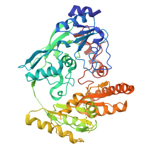
    9QGZ, 9QIT

  • PubMed Abstract: 

    Shotgun metagenomic sequencing has emerged as a powerful tool for exploring microbial diversity and uncovering genes encoding novel biocatalysts from complex environments. Here, we report the discovery and characterization of a new FAD-dependent D-lactate dehydrogenase (PdG-D-LDH) from the gut microbiome of the isopod Porcellio dilatatus . The enzyme was identified through in silico screening using BLAST and AlphaFold3 and functionally characterized as a homodimeric, thermoactive, and thermostable protein, demonstrating the robustness required for biotechnological applications. PdG-D-LDH exhibits a strong catalytic preference toward D-lactate and preferentially reduces quinones over cytochrome c or molecular oxygen. X-ray crystallography revealed a VAO/PCMH-like fold with a solvent-accessible active site that harbors both a FAD cofactor and an Fe(II) ion. Molecular docking studies provided insights into the structural determinants of its stereoselective substrate recognition. Under mild conditions, the enzyme catalyzed the oxidation of D-lactate to pyruvate with a 90% yield after 24 h of reaction, using molecular oxygen as the electron acceptor. This study illustrates how metagenomics, structural biology, and computational tools can jointly drive the discovery of new enzymes with valuable biotechnological applications aligned with circular economic principles. The newly identified D-lactate dehydrogenase, PdG-D-LDH, exhibits thermostability, stereoselectivity, and high catalytic efficiency, providing new insights into the structure-function relationships of lactate-metabolizing enzymes.


  • Organizational Affiliation
    • Instituto de Tecnologia Química e Biológica António Xavier, Universidade Nova de Lisboa, Oeiras, Portugal.

Macromolecule Content 

  • Total Structure Weight: 398.49 kDa 
  • Atom Count: 26,348 
  • Modeled Residue Count: 3,507 
  • Deposited Residue Count: 3,712 
  • Unique protein chains: 1

Macromolecules

Find similar proteins by:|  3D Structure
Entity ID: 1
MoleculeChains  Sequence LengthOrganismDetailsImage
Glycolate oxidase
A, B, C, D, E
A, B, C, D, E, F, G, H
464Porcellio dilatatusMutation(s): 0 
Gene Names: BDW27_113140
UniProt
Find proteins for A0A975LBX7 (Nocardiopsis eucommiae)
Explore A0A975LBX7 
Go to UniProtKB:  A0A975LBX7
Entity Groups
Sequence Clusters30% Identity50% Identity70% Identity90% Identity95% Identity100% Identity
UniProt GroupA0A975LBX7
Sequence Annotations
Expand
Reference Sequence

Small Molecules

Ligands 2 Unique
IDChains Name / Formula / InChI Key2D Diagram3D Interactions
FAD
(Subject of Investigation/LOI)

Query on FAD


Download:Ideal Coordinates CCD File
I [auth A]
K [auth B]
M [auth C]
O [auth D]
Q [auth E]
I [auth A],
K [auth B],
M [auth C],
O [auth D],
Q [auth E],
S [auth F],
U [auth G],
W [auth H]
FLAVIN-ADENINE DINUCLEOTIDE
C27 H33 N9 O15 P2
VWWQXMAJTJZDQX-UYBVJOGSSA-N
FE
(Subject of Investigation/LOI)

Query on FE


Download:Ideal Coordinates CCD File
J [auth A]
L [auth B]
N [auth C]
P [auth D]
R [auth E]
J [auth A],
L [auth B],
N [auth C],
P [auth D],
R [auth E],
T [auth F],
V [auth G],
X [auth H]
FE (III) ION
Fe
VTLYFUHAOXGGBS-UHFFFAOYSA-N

Experimental Data & Validation

Experimental Data

  • Method: X-RAY DIFFRACTION
  • Resolution: 2.84 Å
  • R-Value Free:  0.262 (Depositor) 
  • R-Value Work:  0.207 (Depositor) 
  • R-Value Observed: 0.210 (Depositor) 
Space Group: P 1
Unit Cell:
Length ( Å )Angle ( ˚ )
a = 69.668α = 81.94
b = 103.92β = 77.24
c = 143.827γ = 75.41
Software Package:
Software NamePurpose
PHENIXrefinement
PDB_EXTRACTdata extraction
autoPROCdata reduction
autoPROCdata scaling
PHASERphasing

Structure Validation

View Full Validation Report


Ligand Structure Quality Assessment 


Entry History 

& Funding Information

Deposition Data


Funding OrganizationLocationGrant Number
Foundation for Science and Technology (FCT)Portugal--

Revision History  (Full details and data files)

  • Version 1.0: 2025-12-03
    Type: Initial release
  • Version 1.1: 2025-12-31
    Changes: Database references
  • Version 1.2: 2026-01-14
    Changes: Database references